Zinc »
PDB 5ofn-5onj »
5oj7 »
Zinc in PDB 5oj7: Sirtuin 4 Orthologue From Xenopus Tropicalis in Complex with Adp- Ribose
Protein crystallography data
The structure of Sirtuin 4 Orthologue From Xenopus Tropicalis in Complex with Adp- Ribose, PDB code: 5oj7
was solved by
M.Pannek,
C.Steegborn,
with X-Ray Crystallography technique. A brief refinement statistics is given in the table below:
| Resolution Low / High (Å) | 19.73 / 1.58 |
| Space group | C 2 2 21 |
| Cell size a, b, c (Å), α, β, γ (°) | 69.390, 74.660, 109.700, 90.00, 90.00, 90.00 |
| R / Rfree (%) | 15.2 / 18.6 |
Zinc Binding Sites:
The binding sites of Zinc atom in the Sirtuin 4 Orthologue From Xenopus Tropicalis in Complex with Adp- Ribose
(pdb code 5oj7). This binding sites where shown within
5.0 Angstroms radius around Zinc atom.
In total only one binding site of Zinc was determined in the Sirtuin 4 Orthologue From Xenopus Tropicalis in Complex with Adp- Ribose, PDB code: 5oj7:
In total only one binding site of Zinc was determined in the Sirtuin 4 Orthologue From Xenopus Tropicalis in Complex with Adp- Ribose, PDB code: 5oj7:
Zinc binding site 1 out of 1 in 5oj7
Go back to
Zinc binding site 1 out
of 1 in the Sirtuin 4 Orthologue From Xenopus Tropicalis in Complex with Adp- Ribose
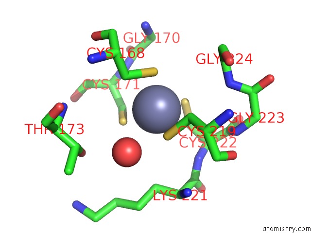
Mono view
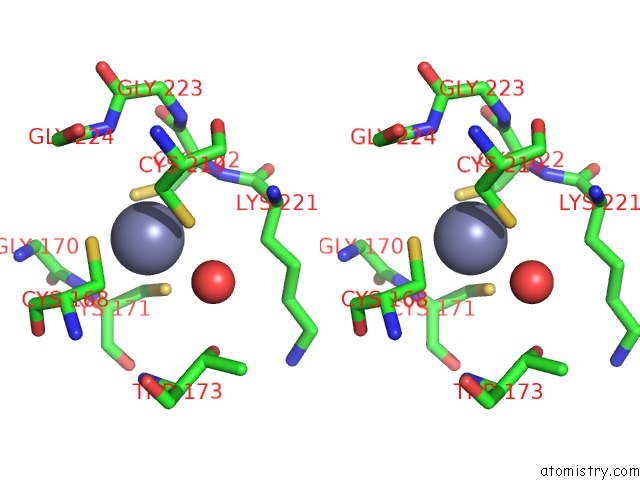
Stereo pair view
Mono view
Stereo pair view
A full contact list of Zinc with other atoms in the Zn binding
site number 1 of Sirtuin 4 Orthologue From Xenopus Tropicalis in Complex with Adp- Ribose within 5.0Å range:
|
Reference:
M.Pannek,
Z.Simic,
M.Fuszard,
M.Meleshin,
D.Rotili,
A.Mai,
M.Schutkowski,
C.Steegborn.
Crystal Structures of the Mitochondrial Deacylase Sirtuin 4 Reveal Isoform-Specific Acyl Recognition and Regulation Features. Nat Commun V. 8 1513 2017.
ISSN: ESSN 2041-1723
PubMed: 29138502
DOI: 10.1038/S41467-017-01701-2
Page generated: Sun Oct 27 23:39:04 2024
ISSN: ESSN 2041-1723
PubMed: 29138502
DOI: 10.1038/S41467-017-01701-2
Last articles
Zn in 9J0NZn in 9J0O
Zn in 9J0P
Zn in 9FJX
Zn in 9EKB
Zn in 9C0F
Zn in 9CAH
Zn in 9CH0
Zn in 9CH3
Zn in 9CH1