Zinc »
PDB 5nf6-5nsp »
5nm9 »
Zinc in PDB 5nm9: Crystal Structure of the Placozoa Trichoplax Adhaerens SMAD4-MH1 Bound to the Ggcgc Site.
Protein crystallography data
The structure of Crystal Structure of the Placozoa Trichoplax Adhaerens SMAD4-MH1 Bound to the Ggcgc Site., PDB code: 5nm9
was solved by
Z.Kaczmarska,
R.Freier,
J.A.Marquez,
M.J.Macias,
with X-Ray Crystallography technique. A brief refinement statistics is given in the table below:
| Resolution Low / High (Å) | 29.46 / 2.43 |
| Space group | P 21 21 21 |
| Cell size a, b, c (Å), α, β, γ (°) | 37.150, 76.980, 145.040, 90.00, 90.00, 90.00 |
| R / Rfree (%) | 22.1 / 25.1 |
Zinc Binding Sites:
The binding sites of Zinc atom in the Crystal Structure of the Placozoa Trichoplax Adhaerens SMAD4-MH1 Bound to the Ggcgc Site.
(pdb code 5nm9). This binding sites where shown within
5.0 Angstroms radius around Zinc atom.
In total 2 binding sites of Zinc where determined in the Crystal Structure of the Placozoa Trichoplax Adhaerens SMAD4-MH1 Bound to the Ggcgc Site., PDB code: 5nm9:
Jump to Zinc binding site number: 1; 2;
In total 2 binding sites of Zinc where determined in the Crystal Structure of the Placozoa Trichoplax Adhaerens SMAD4-MH1 Bound to the Ggcgc Site., PDB code: 5nm9:
Jump to Zinc binding site number: 1; 2;
Zinc binding site 1 out of 2 in 5nm9
Go back to
Zinc binding site 1 out
of 2 in the Crystal Structure of the Placozoa Trichoplax Adhaerens SMAD4-MH1 Bound to the Ggcgc Site.
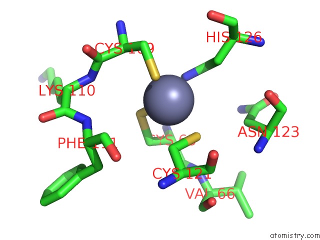
Mono view
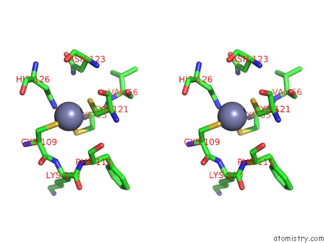
Stereo pair view
Mono view
Stereo pair view
A full contact list of Zinc with other atoms in the Zn binding
site number 1 of Crystal Structure of the Placozoa Trichoplax Adhaerens SMAD4-MH1 Bound to the Ggcgc Site. within 5.0Å range:
|
Zinc binding site 2 out of 2 in 5nm9
Go back to
Zinc binding site 2 out
of 2 in the Crystal Structure of the Placozoa Trichoplax Adhaerens SMAD4-MH1 Bound to the Ggcgc Site.

Mono view
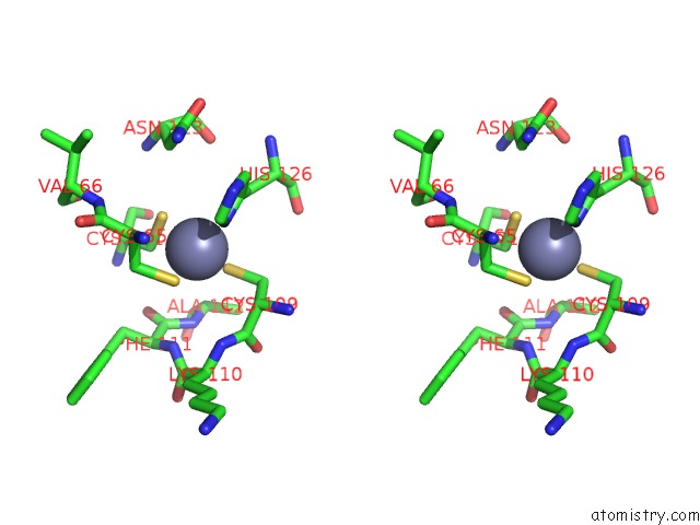
Stereo pair view
Mono view
Stereo pair view
A full contact list of Zinc with other atoms in the Zn binding
site number 2 of Crystal Structure of the Placozoa Trichoplax Adhaerens SMAD4-MH1 Bound to the Ggcgc Site. within 5.0Å range:
|
Reference:
P.Martin-Malpartida,
M.Batet,
Z.Kaczmarska,
R.Freier,
T.Gomes,
E.Aragon,
Y.Zou,
Q.Wang,
Q.Xi,
L.Ruiz,
A.Vea,
J.A.Marquez,
J.Massague,
M.J.Macias.
Structural Basis For Genome Wide Recognition of 5-Bp Gc Motifs By Smad Transcription Factors. Nat Commun V. 8 2070 2017.
ISSN: ESSN 2041-1723
PubMed: 29234012
DOI: 10.1038/S41467-017-02054-6
Page generated: Thu Aug 21 05:39:56 2025
ISSN: ESSN 2041-1723
PubMed: 29234012
DOI: 10.1038/S41467-017-02054-6
Last articles
Zn in 5YI3Zn in 5YIQ
Zn in 5YIX
Zn in 5YI0
Zn in 5YHZ
Zn in 5YHY
Zn in 5YHX
Zn in 5YHT
Zn in 5YEL
Zn in 5YEF