Zinc »
PDB 5yhy-5yo1 »
5yhy »
Zinc in PDB 5yhy: Structure of Lactococcus Lactis Zitr, C30S Mutant
Protein crystallography data
The structure of Structure of Lactococcus Lactis Zitr, C30S Mutant, PDB code: 5yhy
was solved by
Y.Song,
H.Liu,
R.Zhu,
C.Yi,
P.Chen,
with X-Ray Crystallography technique. A brief refinement statistics is given in the table below:
| Resolution Low / High (Å) | 29.22 / 1.65 |
| Space group | P 41 21 2 |
| Cell size a, b, c (Å), α, β, γ (°) | 62.182, 62.182, 117.280, 90.00, 90.00, 90.00 |
| R / Rfree (%) | 20.6 / 22.6 |
Zinc Binding Sites:
The binding sites of Zinc atom in the Structure of Lactococcus Lactis Zitr, C30S Mutant
(pdb code 5yhy). This binding sites where shown within
5.0 Angstroms radius around Zinc atom.
In total only one binding site of Zinc was determined in the Structure of Lactococcus Lactis Zitr, C30S Mutant, PDB code: 5yhy:
In total only one binding site of Zinc was determined in the Structure of Lactococcus Lactis Zitr, C30S Mutant, PDB code: 5yhy:
Zinc binding site 1 out of 1 in 5yhy
Go back to
Zinc binding site 1 out
of 1 in the Structure of Lactococcus Lactis Zitr, C30S Mutant
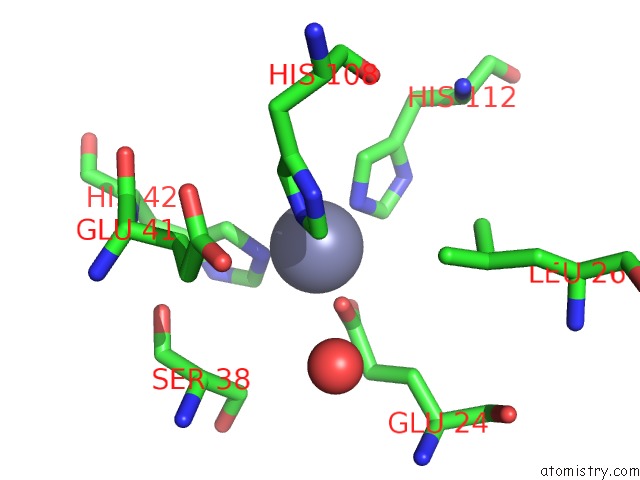
Mono view
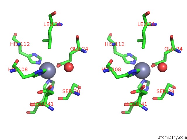
Stereo pair view
Mono view
Stereo pair view
A full contact list of Zinc with other atoms in the Zn binding
site number 1 of Structure of Lactococcus Lactis Zitr, C30S Mutant within 5.0Å range:
|
Reference:
Y.Song,
H.Liu,
R.Zhu,
C.Yi,
P.Chen.
Structure of Lactococcus Lactis Zitr Proc.Natl.Acad.Sci.Usa 2017.
ISSN: ESSN 1091-6490
DOI: 10.1073/PNAS.1708563115
Page generated: Thu Aug 21 11:30:03 2025
ISSN: ESSN 1091-6490
DOI: 10.1073/PNAS.1708563115
Last articles
Zn in 6GYLZn in 6GYK
Zn in 6GXW
Zn in 6GXU
Zn in 6GXA
Zn in 6GXQ
Zn in 6GX3
Zn in 6GXE
Zn in 6GXB
Zn in 6GWU