Zinc »
PDB 5n34-5nek »
5nbc »
Zinc in PDB 5nbc: Structure of Prokaryotic Transcription Factors
Protein crystallography data
The structure of Structure of Prokaryotic Transcription Factors, PDB code: 5nbc
was solved by
J.Perard,
P.Carpentier,
I.Michaud-Soret,
C.Cavazza,
with X-Ray Crystallography technique. A brief refinement statistics is given in the table below:
| Resolution Low / High (Å) | 42.08 / 1.70 |
| Space group | P 1 21 1 |
| Cell size a, b, c (Å), α, β, γ (°) | 53.150, 90.000, 63.950, 90.00, 93.48, 90.00 |
| R / Rfree (%) | 21.5 / 24.4 |
Other elements in 5nbc:
The structure of Structure of Prokaryotic Transcription Factors also contains other interesting chemical elements:
| Manganese | (Mn) | 4 atoms |
Zinc Binding Sites:
The binding sites of Zinc atom in the Structure of Prokaryotic Transcription Factors
(pdb code 5nbc). This binding sites where shown within
5.0 Angstroms radius around Zinc atom.
In total 4 binding sites of Zinc where determined in the Structure of Prokaryotic Transcription Factors, PDB code: 5nbc:
Jump to Zinc binding site number: 1; 2; 3; 4;
In total 4 binding sites of Zinc where determined in the Structure of Prokaryotic Transcription Factors, PDB code: 5nbc:
Jump to Zinc binding site number: 1; 2; 3; 4;
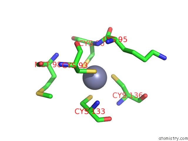
Zinc binding site 1 out of 4 in 5nbc
Go back to
Zinc binding site 1 out
of 4 in the Structure of Prokaryotic Transcription Factors
Mono view

Stereo pair view
Mono view
Stereo pair view
A full contact list of Zinc with other atoms in the Zn binding
site number 1 of Structure of Prokaryotic Transcription Factors within 5.0Å range:
|
Zinc binding site 2 out of 4 in 5nbc
Go back to
Zinc binding site 2 out
of 4 in the Structure of Prokaryotic Transcription Factors

Mono view

Stereo pair view
Mono view
Stereo pair view
A full contact list of Zinc with other atoms in the Zn binding
site number 2 of Structure of Prokaryotic Transcription Factors within 5.0Å range:
|
Zinc binding site 3 out of 4 in 5nbc
Go back to
Zinc binding site 3 out
of 4 in the Structure of Prokaryotic Transcription Factors

Mono view

Stereo pair view
Mono view
Stereo pair view
A full contact list of Zinc with other atoms in the Zn binding
site number 3 of Structure of Prokaryotic Transcription Factors within 5.0Å range:
|
Zinc binding site 4 out of 4 in 5nbc
Go back to
Zinc binding site 4 out
of 4 in the Structure of Prokaryotic Transcription Factors

Mono view

Stereo pair view
Mono view
Stereo pair view
A full contact list of Zinc with other atoms in the Zn binding
site number 4 of Structure of Prokaryotic Transcription Factors within 5.0Å range:
|
Reference:
J.Perard,
S.Nader,
M.Levert,
L.Arnaud,
P.Carpentier,
C.Siebert,
F.Blanquet,
C.Cavazza,
P.Renesto,
D.Schneider,
M.Maurin,
J.Coves,
S.Crouzy,
I.Michaud-Soret.
Structural and Functional Studies of the Metalloregulator Fur Identify A Promoter-Binding Mechanism and Its Role Infrancisella Tularensisvirulence. Commun Biol V. 1 93 2018.
ISSN: ESSN 2399-3642
PubMed: 30271974
DOI: 10.1038/S42003-018-0095-6
Page generated: Sun Oct 27 22:42:31 2024
ISSN: ESSN 2399-3642
PubMed: 30271974
DOI: 10.1038/S42003-018-0095-6
Last articles
Zn in 9J0NZn in 9J0O
Zn in 9J0P
Zn in 9FJX
Zn in 9EKB
Zn in 9C0F
Zn in 9CAH
Zn in 9CH0
Zn in 9CH3
Zn in 9CH1