Zinc »
PDB 5n38-5nel »
5n5g »
Zinc in PDB 5n5g: Crystal Structure of Di-Zinc Metallo-Beta-Lactamase Vim-1
Protein crystallography data
The structure of Crystal Structure of Di-Zinc Metallo-Beta-Lactamase Vim-1, PDB code: 5n5g
was solved by
R.Salimraj,
P.Hinchliffe,
J.Spencer,
with X-Ray Crystallography technique. A brief refinement statistics is given in the table below:
| Resolution Low / High (Å) | 34.25 / 1.29 |
| Space group | P 1 21 1 |
| Cell size a, b, c (Å), α, β, γ (°) | 39.760, 67.940, 40.360, 90.00, 94.01, 90.00 |
| R / Rfree (%) | 14.5 / 15.6 |
Zinc Binding Sites:
The binding sites of Zinc atom in the Crystal Structure of Di-Zinc Metallo-Beta-Lactamase Vim-1
(pdb code 5n5g). This binding sites where shown within
5.0 Angstroms radius around Zinc atom.
In total 3 binding sites of Zinc where determined in the Crystal Structure of Di-Zinc Metallo-Beta-Lactamase Vim-1, PDB code: 5n5g:
Jump to Zinc binding site number: 1; 2; 3;
In total 3 binding sites of Zinc where determined in the Crystal Structure of Di-Zinc Metallo-Beta-Lactamase Vim-1, PDB code: 5n5g:
Jump to Zinc binding site number: 1; 2; 3;
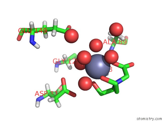
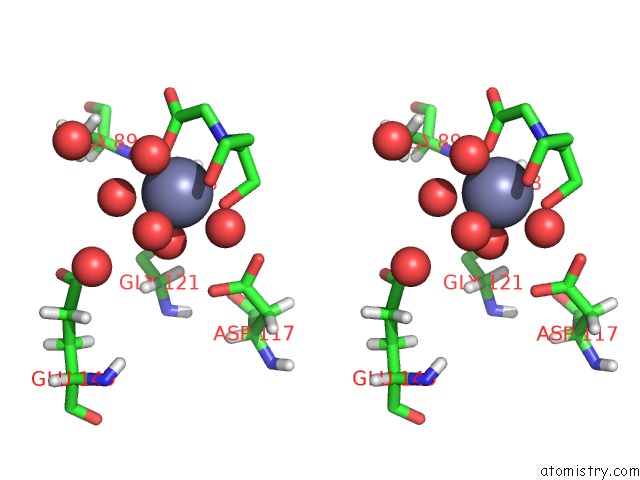
Zinc binding site 1 out of 3 in 5n5g
Go back to
Zinc binding site 1 out
of 3 in the Crystal Structure of Di-Zinc Metallo-Beta-Lactamase Vim-1
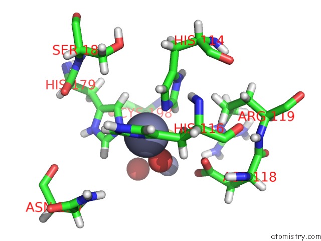
Mono view
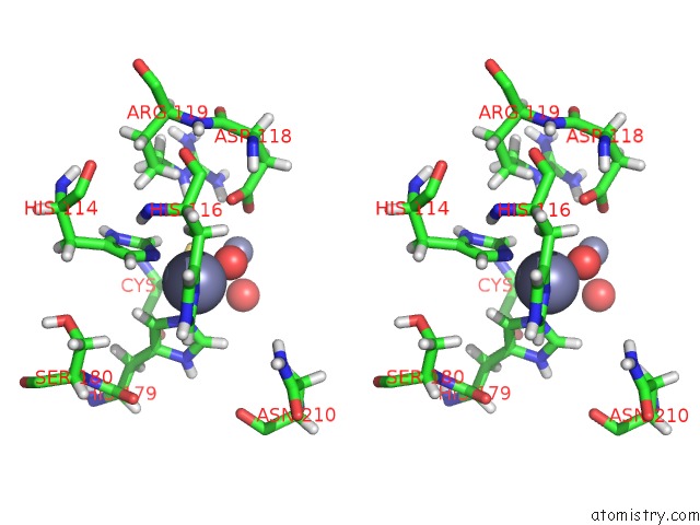
Stereo pair view
Mono view
Stereo pair view
A full contact list of Zinc with other atoms in the Zn binding
site number 1 of Crystal Structure of Di-Zinc Metallo-Beta-Lactamase Vim-1 within 5.0Å range:
|
Zinc binding site 2 out of 3 in 5n5g
Go back to
Zinc binding site 2 out
of 3 in the Crystal Structure of Di-Zinc Metallo-Beta-Lactamase Vim-1
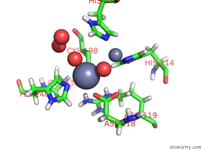
Mono view
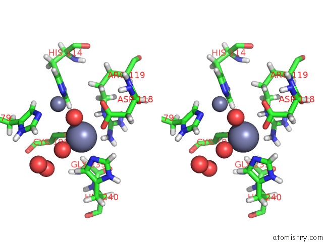
Stereo pair view
Mono view
Stereo pair view
A full contact list of Zinc with other atoms in the Zn binding
site number 2 of Crystal Structure of Di-Zinc Metallo-Beta-Lactamase Vim-1 within 5.0Å range:
|
Zinc binding site 3 out of 3 in 5n5g
Go back to
Zinc binding site 3 out
of 3 in the Crystal Structure of Di-Zinc Metallo-Beta-Lactamase Vim-1
Mono view
Stereo pair view
Mono view
Stereo pair view
A full contact list of Zinc with other atoms in the Zn binding
site number 3 of Crystal Structure of Di-Zinc Metallo-Beta-Lactamase Vim-1 within 5.0Å range:
|
Reference:
R.Salimraj,
P.Hinchliffe,
M.Kosmopoulou,
J.M.Tyrrell,
J.Brem,
S.S.Van Berkel,
A.Verma,
R.J.Owens,
M.A.Mcdonough,
T.R.Walsh,
C.J.Schofield,
J.Spencer.
Crystal Structures of Vim-1 Complexes Explain Active Site Heterogeneity in Vim-Class Metallo-Beta-Lactamases. Febs J. V. 286 169 2019.
ISSN: ISSN 1742-4658
PubMed: 30430727
DOI: 10.1111/FEBS.14695
Page generated: Thu Aug 21 05:27:41 2025
ISSN: ISSN 1742-4658
PubMed: 30430727
DOI: 10.1111/FEBS.14695
Last articles
Zn in 6FTMZn in 6FP6
Zn in 6FT0
Zn in 6FS3
Zn in 6FSO
Zn in 6FSM
Zn in 6FRZ
Zn in 6FRW
Zn in 6FQE
Zn in 6FRD