Zinc »
PDB 6fhq-6ft0 »
6frw »
Zinc in PDB 6frw: X-Ray Structure of the Levansucrase From Erwinia Tasmaniensis
Enzymatic activity of X-Ray Structure of the Levansucrase From Erwinia Tasmaniensis
All present enzymatic activity of X-Ray Structure of the Levansucrase From Erwinia Tasmaniensis:
2.4.1.10;
2.4.1.10;
Protein crystallography data
The structure of X-Ray Structure of the Levansucrase From Erwinia Tasmaniensis, PDB code: 6frw
was solved by
I.Polsinelli,
M.Salomone-Stagni,
R.Caliandro,
N.Demitri,
S.Benini,
with X-Ray Crystallography technique. A brief refinement statistics is given in the table below:
| Resolution Low / High (Å) | 41.15 / 1.52 |
| Space group | P 41 21 2 |
| Cell size a, b, c (Å), α, β, γ (°) | 128.493, 128.493, 58.945, 90.00, 90.00, 90.00 |
| R / Rfree (%) | 17.9 / 20.9 |
Zinc Binding Sites:
The binding sites of Zinc atom in the X-Ray Structure of the Levansucrase From Erwinia Tasmaniensis
(pdb code 6frw). This binding sites where shown within
5.0 Angstroms radius around Zinc atom.
In total 3 binding sites of Zinc where determined in the X-Ray Structure of the Levansucrase From Erwinia Tasmaniensis, PDB code: 6frw:
Jump to Zinc binding site number: 1; 2; 3;
In total 3 binding sites of Zinc where determined in the X-Ray Structure of the Levansucrase From Erwinia Tasmaniensis, PDB code: 6frw:
Jump to Zinc binding site number: 1; 2; 3;
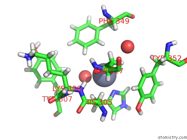
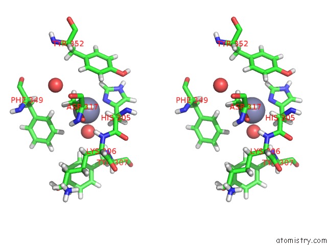
Zinc binding site 1 out of 3 in 6frw
Go back to
Zinc binding site 1 out
of 3 in the X-Ray Structure of the Levansucrase From Erwinia Tasmaniensis
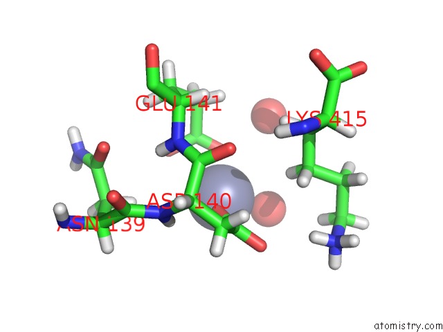
Mono view
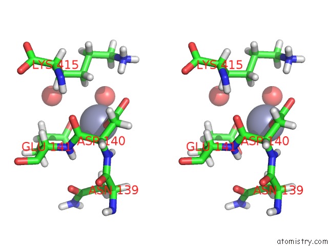
Stereo pair view
Mono view
Stereo pair view
A full contact list of Zinc with other atoms in the Zn binding
site number 1 of X-Ray Structure of the Levansucrase From Erwinia Tasmaniensis within 5.0Å range:
|
Zinc binding site 2 out of 3 in 6frw
Go back to
Zinc binding site 2 out
of 3 in the X-Ray Structure of the Levansucrase From Erwinia Tasmaniensis
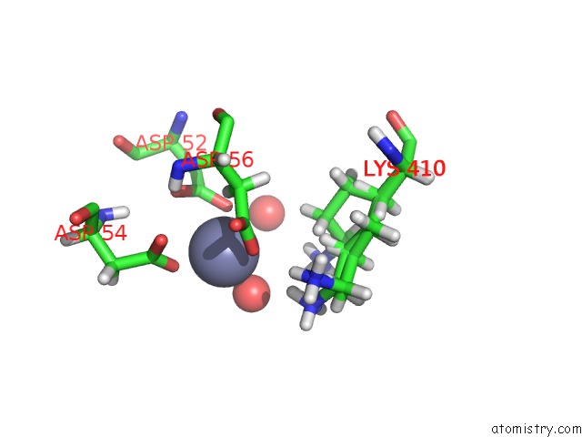
Mono view
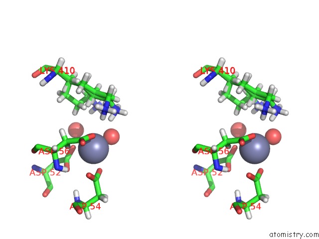
Stereo pair view
Mono view
Stereo pair view
A full contact list of Zinc with other atoms in the Zn binding
site number 2 of X-Ray Structure of the Levansucrase From Erwinia Tasmaniensis within 5.0Å range:
|
Zinc binding site 3 out of 3 in 6frw
Go back to
Zinc binding site 3 out
of 3 in the X-Ray Structure of the Levansucrase From Erwinia Tasmaniensis
Mono view
Stereo pair view
Mono view
Stereo pair view
A full contact list of Zinc with other atoms in the Zn binding
site number 3 of X-Ray Structure of the Levansucrase From Erwinia Tasmaniensis within 5.0Å range:
|
Reference:
I.Polsinelli,
R.Caliandro,
M.Salomone-Stagni,
N.Demitri,
M.Rejzek,
R.A.Field,
S.Benini.
Comparison of the Levansucrase From the Epiphyte Erwinia Tasmaniensis Vs Its Homologue From the Phytopathogen Erwinia Amylovora. Int. J. Biol. Macromol. V. 127 496 2019.
ISSN: ISSN 1879-0003
PubMed: 30660564
DOI: 10.1016/J.IJBIOMAC.2019.01.074
Page generated: Thu Aug 21 14:24:43 2025
ISSN: ISSN 1879-0003
PubMed: 30660564
DOI: 10.1016/J.IJBIOMAC.2019.01.074
Last articles
Zn in 6P2KZn in 6P3Z
Zn in 6P3Y
Zn in 6P3X
Zn in 6P0R
Zn in 6P2T
Zn in 6P1Z
Zn in 6P1K
Zn in 6P20
Zn in 6P1G