Zinc »
PDB 5mcw-5msl »
5mkw »
Zinc in PDB 5mkw: Crystal Structure of the Human ZRANB3 Hnh Domain
Protein crystallography data
The structure of Crystal Structure of the Human ZRANB3 Hnh Domain, PDB code: 5mkw
was solved by
A.Ariza,
with X-Ray Crystallography technique. A brief refinement statistics is given in the table below:
| Resolution Low / High (Å) | 54.48 / 2.00 |
| Space group | P 21 21 21 |
| Cell size a, b, c (Å), α, β, γ (°) | 56.841, 67.552, 92.161, 90.00, 90.00, 90.00 |
| R / Rfree (%) | 19.2 / 23.6 |
Zinc Binding Sites:
The binding sites of Zinc atom in the Crystal Structure of the Human ZRANB3 Hnh Domain
(pdb code 5mkw). This binding sites where shown within
5.0 Angstroms radius around Zinc atom.
In total 2 binding sites of Zinc where determined in the Crystal Structure of the Human ZRANB3 Hnh Domain, PDB code: 5mkw:
Jump to Zinc binding site number: 1; 2;
In total 2 binding sites of Zinc where determined in the Crystal Structure of the Human ZRANB3 Hnh Domain, PDB code: 5mkw:
Jump to Zinc binding site number: 1; 2;
Zinc binding site 1 out of 2 in 5mkw
Go back to
Zinc binding site 1 out
of 2 in the Crystal Structure of the Human ZRANB3 Hnh Domain
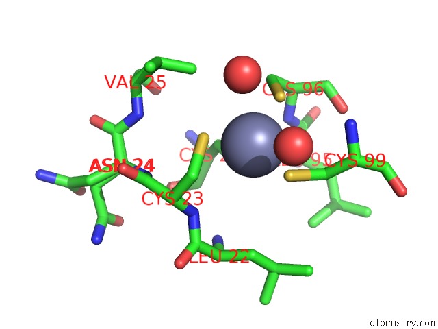
Mono view
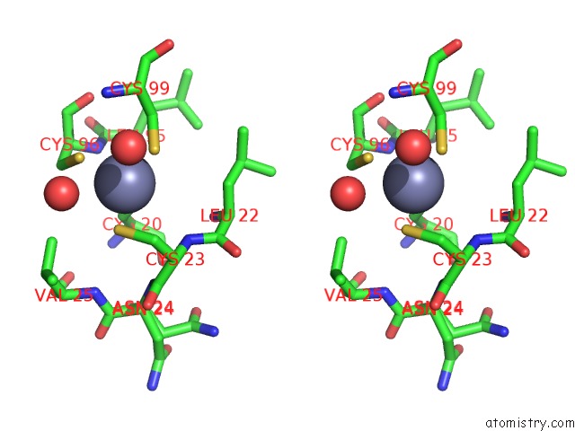
Stereo pair view
Mono view
Stereo pair view
A full contact list of Zinc with other atoms in the Zn binding
site number 1 of Crystal Structure of the Human ZRANB3 Hnh Domain within 5.0Å range:
|
Zinc binding site 2 out of 2 in 5mkw
Go back to
Zinc binding site 2 out
of 2 in the Crystal Structure of the Human ZRANB3 Hnh Domain
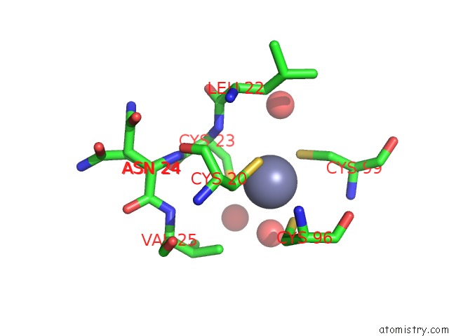
Mono view
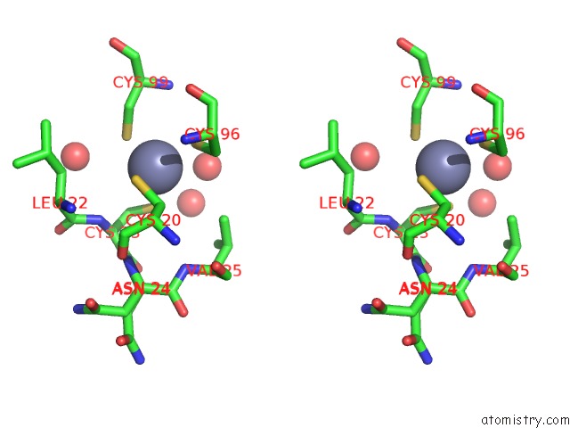
Stereo pair view
Mono view
Stereo pair view
A full contact list of Zinc with other atoms in the Zn binding
site number 2 of Crystal Structure of the Human ZRANB3 Hnh Domain within 5.0Å range:
|
Reference:
M.Sebesta,
C.D.O.Cooper,
A.Ariza,
C.J.Carnie,
D.Ahel.
Structural Insights Into the Function of ZRANB3 in Replication Stress Response. Nat Commun V. 8 15847 2017.
ISSN: ESSN 2041-1723
PubMed: 28621305
DOI: 10.1038/NCOMMS15847
Page generated: Sun Oct 27 22:09:59 2024
ISSN: ESSN 2041-1723
PubMed: 28621305
DOI: 10.1038/NCOMMS15847
Last articles
Zn in 9J0NZn in 9J0O
Zn in 9J0P
Zn in 9FJX
Zn in 9EKB
Zn in 9C0F
Zn in 9CAH
Zn in 9CH0
Zn in 9CH3
Zn in 9CH1