Zinc »
PDB 5g2u-5gl7 »
5giv »
Zinc in PDB 5giv: Crystal Structure of M32 Carboxypeptidase From Deinococcus Radiodurans R1
Enzymatic activity of Crystal Structure of M32 Carboxypeptidase From Deinococcus Radiodurans R1
All present enzymatic activity of Crystal Structure of M32 Carboxypeptidase From Deinococcus Radiodurans R1:
3.4.17.19;
3.4.17.19;
Protein crystallography data
The structure of Crystal Structure of M32 Carboxypeptidase From Deinococcus Radiodurans R1, PDB code: 5giv
was solved by
B.Sharma,
R.Singh,
P.Yadav,
B.Ghosh,
A.Kumar,
S.N.Jamdar,
R.D.Makde,
with X-Ray Crystallography technique. A brief refinement statistics is given in the table below:
| Resolution Low / High (Å) | 47.97 / 2.40 |
| Space group | C 2 2 21 |
| Cell size a, b, c (Å), α, β, γ (°) | 134.857, 256.632, 199.214, 90.00, 90.00, 90.00 |
| R / Rfree (%) | 20.6 / 24.3 |
Zinc Binding Sites:
The binding sites of Zinc atom in the Crystal Structure of M32 Carboxypeptidase From Deinococcus Radiodurans R1
(pdb code 5giv). This binding sites where shown within
5.0 Angstroms radius around Zinc atom.
In total 6 binding sites of Zinc where determined in the Crystal Structure of M32 Carboxypeptidase From Deinococcus Radiodurans R1, PDB code: 5giv:
Jump to Zinc binding site number: 1; 2; 3; 4; 5; 6;
In total 6 binding sites of Zinc where determined in the Crystal Structure of M32 Carboxypeptidase From Deinococcus Radiodurans R1, PDB code: 5giv:
Jump to Zinc binding site number: 1; 2; 3; 4; 5; 6;
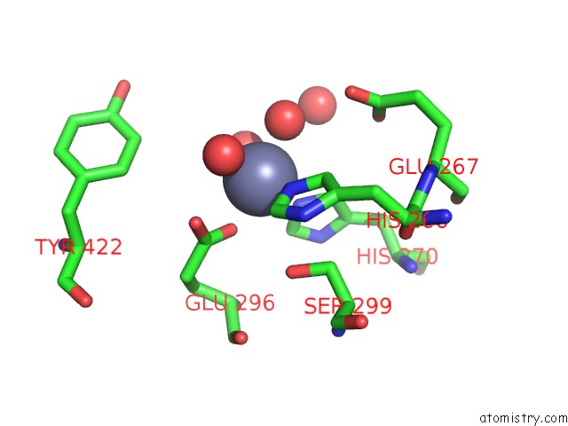
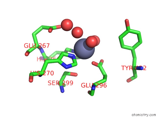
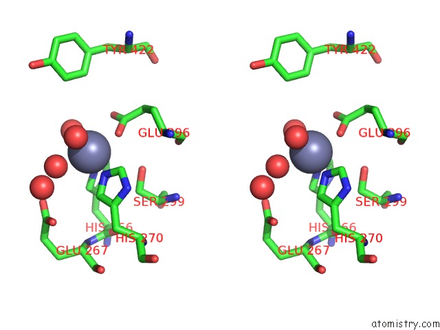
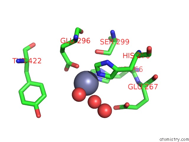
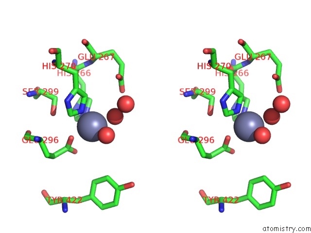
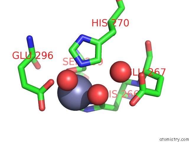
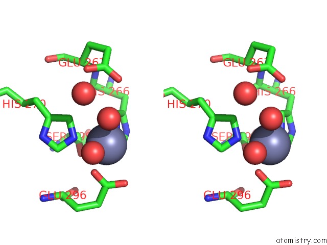
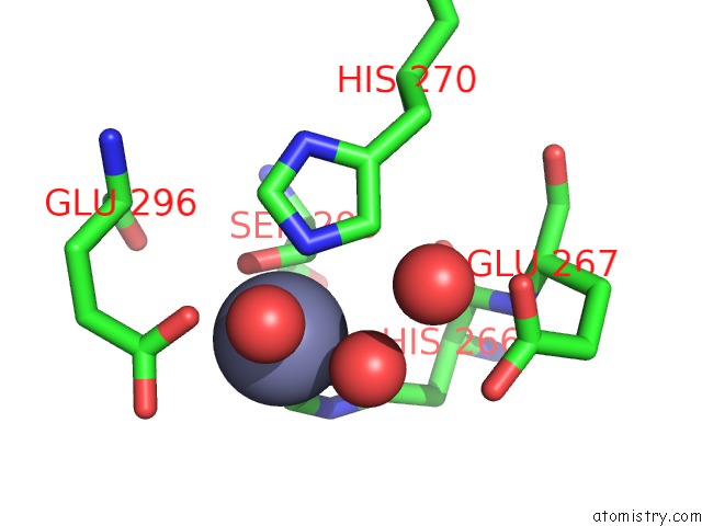
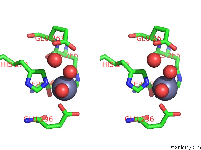
Zinc binding site 1 out of 6 in 5giv
Go back to
Zinc binding site 1 out
of 6 in the Crystal Structure of M32 Carboxypeptidase From Deinococcus Radiodurans R1
Mono view
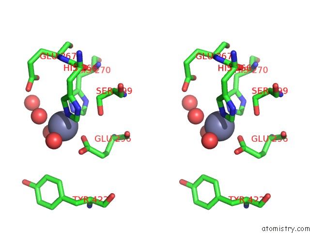
Stereo pair view
Mono view
Stereo pair view
A full contact list of Zinc with other atoms in the Zn binding
site number 1 of Crystal Structure of M32 Carboxypeptidase From Deinococcus Radiodurans R1 within 5.0Å range:
|
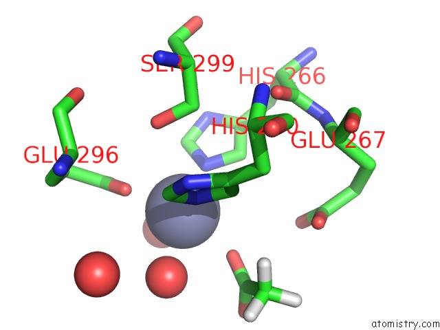
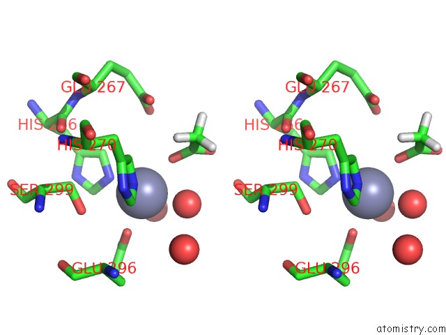
Zinc binding site 2 out of 6 in 5giv
Go back to
Zinc binding site 2 out
of 6 in the Crystal Structure of M32 Carboxypeptidase From Deinococcus Radiodurans R1
Mono view
Stereo pair view
Mono view
Stereo pair view
A full contact list of Zinc with other atoms in the Zn binding
site number 2 of Crystal Structure of M32 Carboxypeptidase From Deinococcus Radiodurans R1 within 5.0Å range:
|
Zinc binding site 3 out of 6 in 5giv
Go back to
Zinc binding site 3 out
of 6 in the Crystal Structure of M32 Carboxypeptidase From Deinococcus Radiodurans R1
Mono view
Stereo pair view
Mono view
Stereo pair view
A full contact list of Zinc with other atoms in the Zn binding
site number 3 of Crystal Structure of M32 Carboxypeptidase From Deinococcus Radiodurans R1 within 5.0Å range:
|
Zinc binding site 4 out of 6 in 5giv
Go back to
Zinc binding site 4 out
of 6 in the Crystal Structure of M32 Carboxypeptidase From Deinococcus Radiodurans R1
Mono view
Stereo pair view
Mono view
Stereo pair view
A full contact list of Zinc with other atoms in the Zn binding
site number 4 of Crystal Structure of M32 Carboxypeptidase From Deinococcus Radiodurans R1 within 5.0Å range:
|
Zinc binding site 5 out of 6 in 5giv
Go back to
Zinc binding site 5 out
of 6 in the Crystal Structure of M32 Carboxypeptidase From Deinococcus Radiodurans R1
Mono view
Stereo pair view
Mono view
Stereo pair view
A full contact list of Zinc with other atoms in the Zn binding
site number 5 of Crystal Structure of M32 Carboxypeptidase From Deinococcus Radiodurans R1 within 5.0Å range:
|
Zinc binding site 6 out of 6 in 5giv
Go back to
Zinc binding site 6 out
of 6 in the Crystal Structure of M32 Carboxypeptidase From Deinococcus Radiodurans R1
Mono view
Stereo pair view
Mono view
Stereo pair view
A full contact list of Zinc with other atoms in the Zn binding
site number 6 of Crystal Structure of M32 Carboxypeptidase From Deinococcus Radiodurans R1 within 5.0Å range:
|
Reference:
B.Sharma,
S.N.Jamdar,
B.Ghosh,
P.Yadav,
A.Kumar,
S.Kundu,
V.D.Goyal,
R.D.Makde.
Active Site Gate of M32 Carboxypeptidases Illuminated By Crystal Structure and Molecular Dynamics Simulations Biochim. Biophys. Acta V.1865 1406 2017.
ISSN: ISSN 0006-3002
PubMed: 28844748
DOI: 10.1016/J.BBAPAP.2017.07.023
Page generated: Sun Oct 27 17:00:25 2024
ISSN: ISSN 0006-3002
PubMed: 28844748
DOI: 10.1016/J.BBAPAP.2017.07.023
Last articles
Zn in 9J0NZn in 9J0O
Zn in 9J0P
Zn in 9FJX
Zn in 9EKB
Zn in 9C0F
Zn in 9CAH
Zn in 9CH0
Zn in 9CH3
Zn in 9CH1