Zinc »
PDB 5g2u-5gl7 »
5g32 »
Zinc in PDB 5g32: Structure of RAD14 in Complex with Acetylaminophenyl-Guanine Containing Dna
Protein crystallography data
The structure of Structure of RAD14 in Complex with Acetylaminophenyl-Guanine Containing Dna, PDB code: 5g32
was solved by
N.Simon,
C.Ebert,
S.Schneider,
with X-Ray Crystallography technique. A brief refinement statistics is given in the table below:
| Resolution Low / High (Å) | 52.96 / 2.20 |
| Space group | P 41 |
| Cell size a, b, c (Å), α, β, γ (°) | 52.964, 52.964, 130.945, 90.00, 90.00, 90.00 |
| R / Rfree (%) | 21.8 / 26.6 |
Zinc Binding Sites:
The binding sites of Zinc atom in the Structure of RAD14 in Complex with Acetylaminophenyl-Guanine Containing Dna
(pdb code 5g32). This binding sites where shown within
5.0 Angstroms radius around Zinc atom.
In total 2 binding sites of Zinc where determined in the Structure of RAD14 in Complex with Acetylaminophenyl-Guanine Containing Dna, PDB code: 5g32:
Jump to Zinc binding site number: 1; 2;
In total 2 binding sites of Zinc where determined in the Structure of RAD14 in Complex with Acetylaminophenyl-Guanine Containing Dna, PDB code: 5g32:
Jump to Zinc binding site number: 1; 2;
Zinc binding site 1 out of 2 in 5g32
Go back to
Zinc binding site 1 out
of 2 in the Structure of RAD14 in Complex with Acetylaminophenyl-Guanine Containing Dna
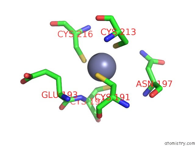
Mono view
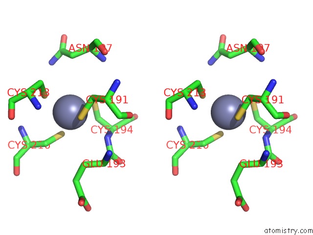
Stereo pair view
Mono view
Stereo pair view
A full contact list of Zinc with other atoms in the Zn binding
site number 1 of Structure of RAD14 in Complex with Acetylaminophenyl-Guanine Containing Dna within 5.0Å range:
|
Zinc binding site 2 out of 2 in 5g32
Go back to
Zinc binding site 2 out
of 2 in the Structure of RAD14 in Complex with Acetylaminophenyl-Guanine Containing Dna
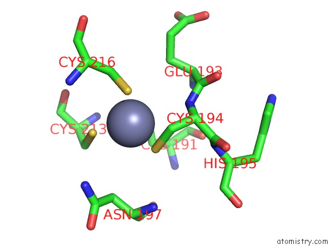
Mono view
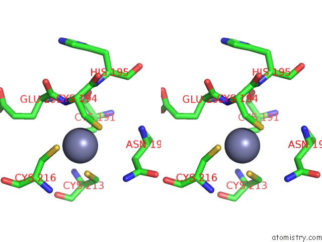
Stereo pair view
Mono view
Stereo pair view
A full contact list of Zinc with other atoms in the Zn binding
site number 2 of Structure of RAD14 in Complex with Acetylaminophenyl-Guanine Containing Dna within 5.0Å range:
|
Reference:
S.Schneider,
N.Simon,
C.Ebert.
Structural Basis For Bulky Adduct Dna Lesion Recognition By the Nucleotide Excision Repair Protein RAD14. Chemistry V. 22 10782 2016.
ISSN: ISSN 0947-6539
PubMed: 27223336
DOI: 10.1002/CHEM.201602438
Page generated: Sun Oct 27 16:53:54 2024
ISSN: ISSN 0947-6539
PubMed: 27223336
DOI: 10.1002/CHEM.201602438
Last articles
Zn in 9J0NZn in 9J0O
Zn in 9J0P
Zn in 9FJX
Zn in 9EKB
Zn in 9C0F
Zn in 9CAH
Zn in 9CH0
Zn in 9CH3
Zn in 9CH1