Zinc »
PDB 5g2u-5gl7 »
5g31 »
Zinc in PDB 5g31: Crystallographic Structure of Mutant C73S of Thioredoxin From Litopenaeus Vannamei
Enzymatic activity of Crystallographic Structure of Mutant C73S of Thioredoxin From Litopenaeus Vannamei
All present enzymatic activity of Crystallographic Structure of Mutant C73S of Thioredoxin From Litopenaeus Vannamei:
1.8.1.9;
1.8.1.9;
Protein crystallography data
The structure of Crystallographic Structure of Mutant C73S of Thioredoxin From Litopenaeus Vannamei, PDB code: 5g31
was solved by
A.A.Campos-Acevedo,
E.Rudino-Pinera,
with X-Ray Crystallography technique. A brief refinement statistics is given in the table below:
| Resolution Low / High (Å) | 44.86 / 2.00 |
| Space group | P 42 21 2 |
| Cell size a, b, c (Å), α, β, γ (°) | 63.444, 63.444, 56.297, 90.00, 90.00, 90.00 |
| R / Rfree (%) | 20.8 / 23.9 |
Zinc Binding Sites:
The binding sites of Zinc atom in the Crystallographic Structure of Mutant C73S of Thioredoxin From Litopenaeus Vannamei
(pdb code 5g31). This binding sites where shown within
5.0 Angstroms radius around Zinc atom.
In total only one binding site of Zinc was determined in the Crystallographic Structure of Mutant C73S of Thioredoxin From Litopenaeus Vannamei, PDB code: 5g31:
In total only one binding site of Zinc was determined in the Crystallographic Structure of Mutant C73S of Thioredoxin From Litopenaeus Vannamei, PDB code: 5g31:
Zinc binding site 1 out of 1 in 5g31
Go back to
Zinc binding site 1 out
of 1 in the Crystallographic Structure of Mutant C73S of Thioredoxin From Litopenaeus Vannamei
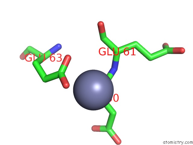
Mono view
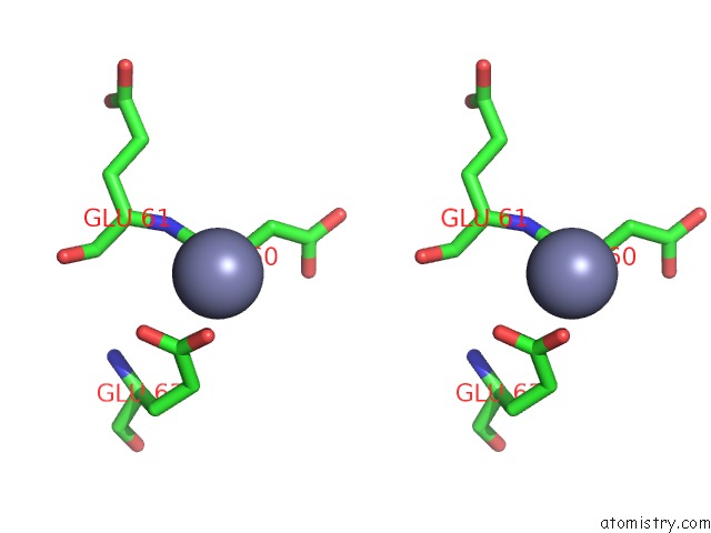
Stereo pair view
Mono view
Stereo pair view
A full contact list of Zinc with other atoms in the Zn binding
site number 1 of Crystallographic Structure of Mutant C73S of Thioredoxin From Litopenaeus Vannamei within 5.0Å range:
|
Reference:
A.A.Campos-Acevedo,
R.R.Sotelo-Mundo,
J.Perez,
E.Rudino-Pinera.
Is Dimerization A Common Feature in Thioredoxins? the Case of Thioredoxin From Litopenaeus Vannamei. Acta Crystallogr D Struct V. 73 326 2017BIOL.
ISSN: ISSN 2059-7983
PubMed: 28375144
DOI: 10.1107/S2059798317002066
Page generated: Sun Oct 27 16:53:54 2024
ISSN: ISSN 2059-7983
PubMed: 28375144
DOI: 10.1107/S2059798317002066
Last articles
Zn in 9J0NZn in 9J0O
Zn in 9J0P
Zn in 9FJX
Zn in 9EKB
Zn in 9C0F
Zn in 9CAH
Zn in 9CH0
Zn in 9CH3
Zn in 9CH1