Zinc »
PDB 5drr-5e9a »
5e0y »
Zinc in PDB 5e0y: Crystal Structure of Pasta Domain 4 of Mycobacterium Tuberculosis Protein Kinase B
Enzymatic activity of Crystal Structure of Pasta Domain 4 of Mycobacterium Tuberculosis Protein Kinase B
All present enzymatic activity of Crystal Structure of Pasta Domain 4 of Mycobacterium Tuberculosis Protein Kinase B:
2.7.11.1;
2.7.11.1;
Protein crystallography data
The structure of Crystal Structure of Pasta Domain 4 of Mycobacterium Tuberculosis Protein Kinase B, PDB code: 5e0y
was solved by
D.M.Prigozhin,
Tb Structural Genomics Consortium (Tbsgc),
with X-Ray Crystallography technique. A brief refinement statistics is given in the table below:
| Resolution Low / High (Å) | 34.61 / 2.00 |
| Space group | P 65 2 2 |
| Cell size a, b, c (Å), α, β, γ (°) | 41.658, 41.658, 122.517, 90.00, 90.00, 120.00 |
| R / Rfree (%) | 21.5 / 26.4 |
Zinc Binding Sites:
The binding sites of Zinc atom in the Crystal Structure of Pasta Domain 4 of Mycobacterium Tuberculosis Protein Kinase B
(pdb code 5e0y). This binding sites where shown within
5.0 Angstroms radius around Zinc atom.
In total 9 binding sites of Zinc where determined in the Crystal Structure of Pasta Domain 4 of Mycobacterium Tuberculosis Protein Kinase B, PDB code: 5e0y:
Jump to Zinc binding site number: 1; 2; 3; 4; 5; 6; 7; 8; 9;
In total 9 binding sites of Zinc where determined in the Crystal Structure of Pasta Domain 4 of Mycobacterium Tuberculosis Protein Kinase B, PDB code: 5e0y:
Jump to Zinc binding site number: 1; 2; 3; 4; 5; 6; 7; 8; 9;
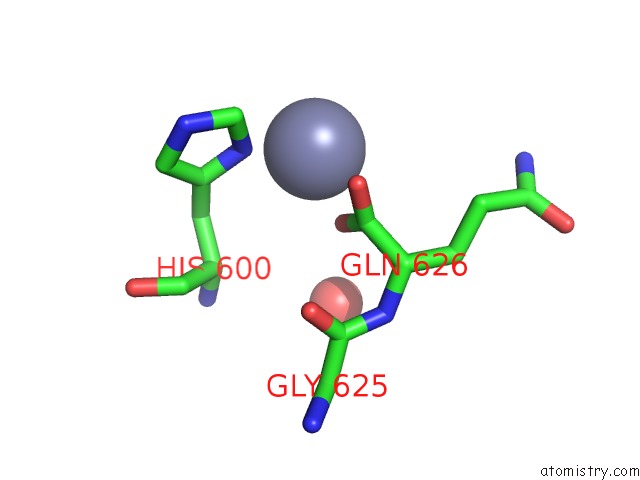
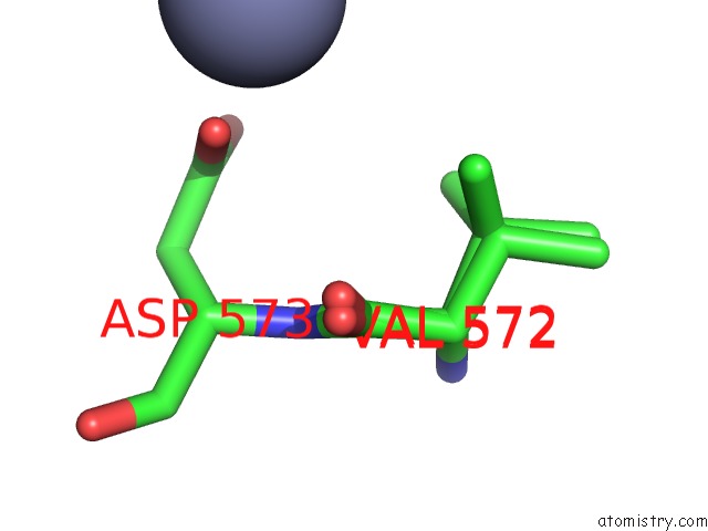
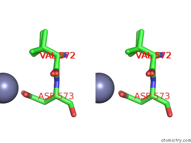
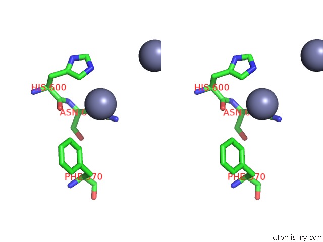
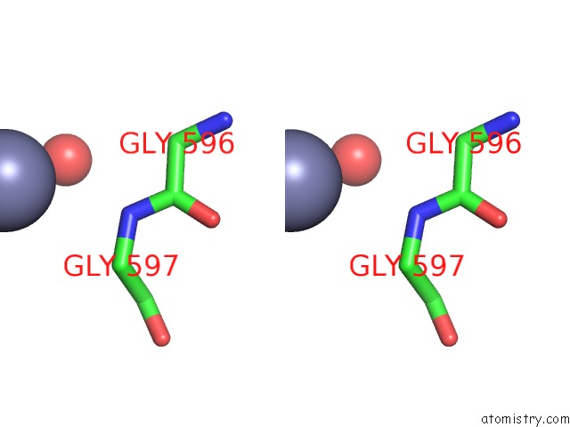
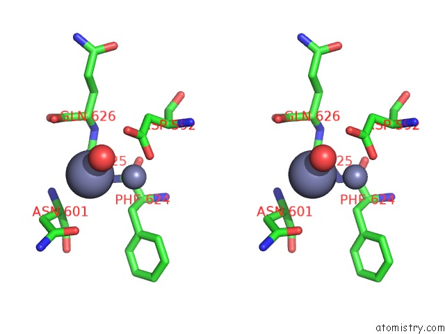
Zinc binding site 1 out of 9 in 5e0y
Go back to
Zinc binding site 1 out
of 9 in the Crystal Structure of Pasta Domain 4 of Mycobacterium Tuberculosis Protein Kinase B
Mono view
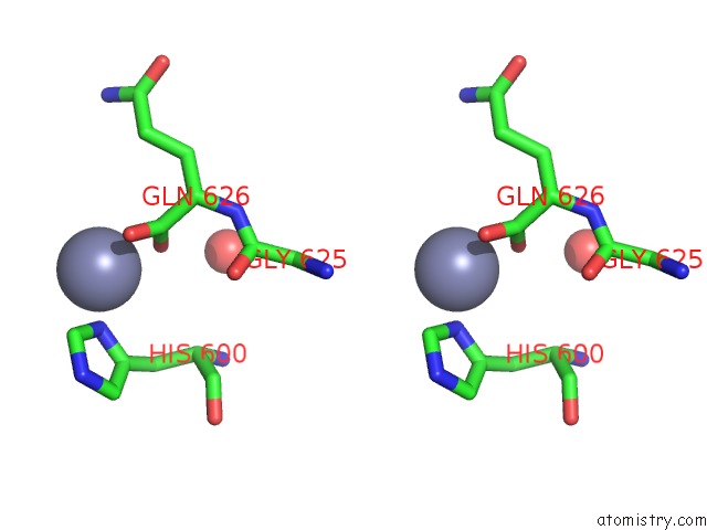
Stereo pair view
Mono view
Stereo pair view
A full contact list of Zinc with other atoms in the Zn binding
site number 1 of Crystal Structure of Pasta Domain 4 of Mycobacterium Tuberculosis Protein Kinase B within 5.0Å range:
|
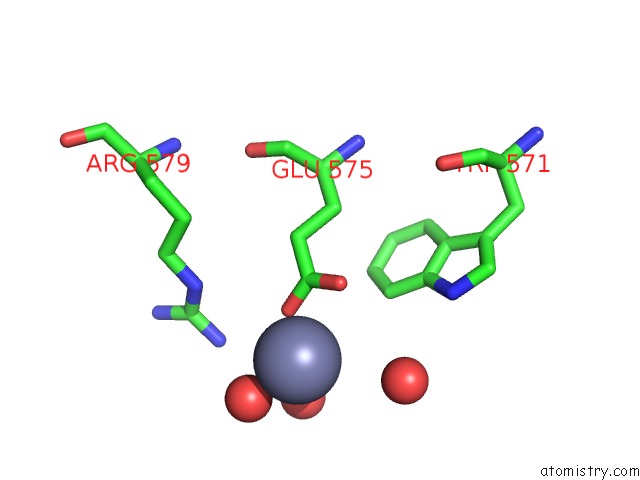
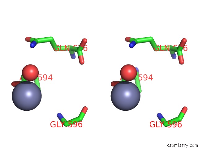
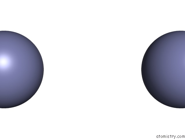
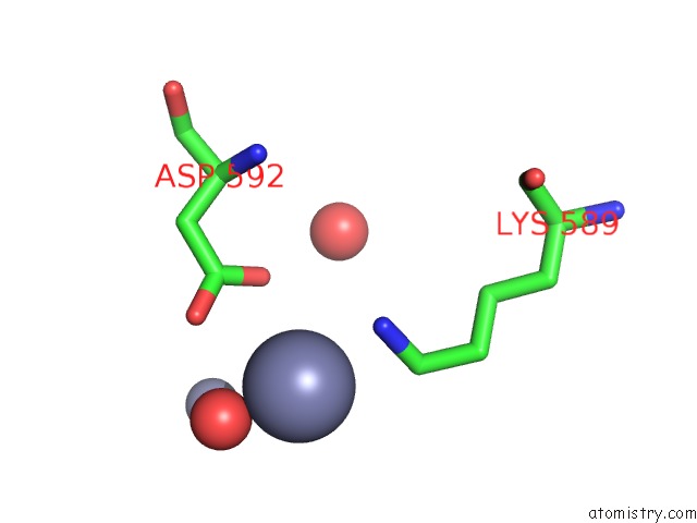
Zinc binding site 2 out of 9 in 5e0y
Go back to
Zinc binding site 2 out
of 9 in the Crystal Structure of Pasta Domain 4 of Mycobacterium Tuberculosis Protein Kinase B
Mono view
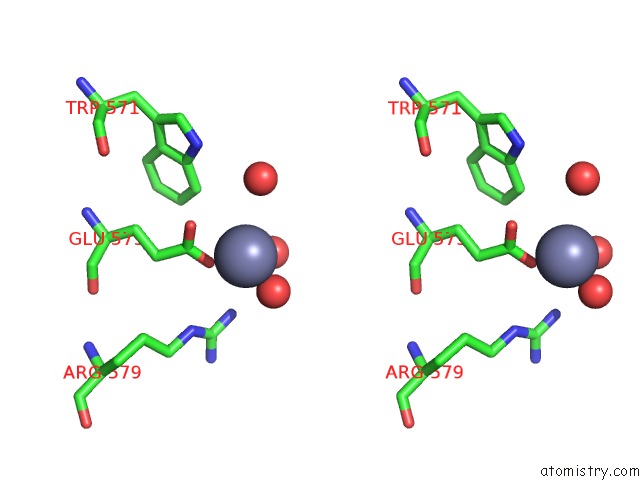
Stereo pair view
Mono view
Stereo pair view
A full contact list of Zinc with other atoms in the Zn binding
site number 2 of Crystal Structure of Pasta Domain 4 of Mycobacterium Tuberculosis Protein Kinase B within 5.0Å range:
|
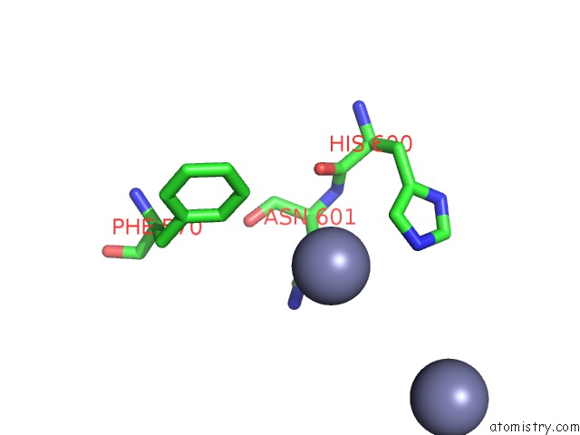
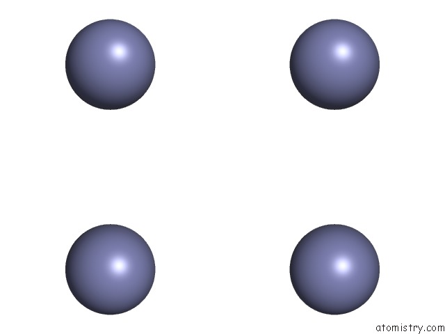
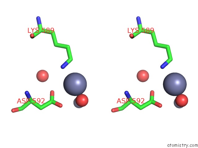
Zinc binding site 3 out of 9 in 5e0y
Go back to
Zinc binding site 3 out
of 9 in the Crystal Structure of Pasta Domain 4 of Mycobacterium Tuberculosis Protein Kinase B
Mono view
Stereo pair view
Mono view
Stereo pair view
A full contact list of Zinc with other atoms in the Zn binding
site number 3 of Crystal Structure of Pasta Domain 4 of Mycobacterium Tuberculosis Protein Kinase B within 5.0Å range:
|
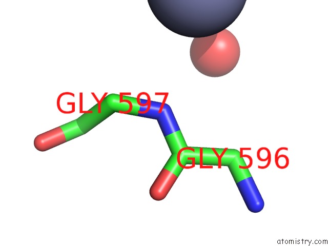
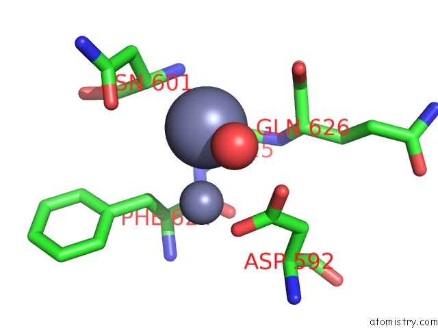
Zinc binding site 4 out of 9 in 5e0y
Go back to
Zinc binding site 4 out
of 9 in the Crystal Structure of Pasta Domain 4 of Mycobacterium Tuberculosis Protein Kinase B

Mono view
Stereo pair view
Mono view
Stereo pair view
A full contact list of Zinc with other atoms in the Zn binding
site number 4 of Crystal Structure of Pasta Domain 4 of Mycobacterium Tuberculosis Protein Kinase B within 5.0Å range:
|
Zinc binding site 5 out of 9 in 5e0y
Go back to
Zinc binding site 5 out
of 9 in the Crystal Structure of Pasta Domain 4 of Mycobacterium Tuberculosis Protein Kinase B
Mono view
Stereo pair view
Mono view
Stereo pair view
A full contact list of Zinc with other atoms in the Zn binding
site number 5 of Crystal Structure of Pasta Domain 4 of Mycobacterium Tuberculosis Protein Kinase B within 5.0Å range:
|
Zinc binding site 6 out of 9 in 5e0y
Go back to
Zinc binding site 6 out
of 9 in the Crystal Structure of Pasta Domain 4 of Mycobacterium Tuberculosis Protein Kinase B
Mono view
Stereo pair view
Mono view
Stereo pair view
| A full contact list of Zinc with other atoms in the Zn binding site number 6 of Crystal Structure of Pasta Domain 4 of Mycobacterium Tuberculosis Protein Kinase B within 5.0Å range: |
Zinc binding site 7 out of 9 in 5e0y
Go back to
Zinc binding site 7 out
of 9 in the Crystal Structure of Pasta Domain 4 of Mycobacterium Tuberculosis Protein Kinase B
Mono view
Stereo pair view
Mono view
Stereo pair view
A full contact list of Zinc with other atoms in the Zn binding
site number 7 of Crystal Structure of Pasta Domain 4 of Mycobacterium Tuberculosis Protein Kinase B within 5.0Å range:
|
Zinc binding site 8 out of 9 in 5e0y
Go back to
Zinc binding site 8 out
of 9 in the Crystal Structure of Pasta Domain 4 of Mycobacterium Tuberculosis Protein Kinase B
Mono view
Stereo pair view
Mono view
Stereo pair view
A full contact list of Zinc with other atoms in the Zn binding
site number 8 of Crystal Structure of Pasta Domain 4 of Mycobacterium Tuberculosis Protein Kinase B within 5.0Å range:
|
Zinc binding site 9 out of 9 in 5e0y
Go back to
Zinc binding site 9 out
of 9 in the Crystal Structure of Pasta Domain 4 of Mycobacterium Tuberculosis Protein Kinase B
Mono view
Stereo pair view
Mono view
Stereo pair view
A full contact list of Zinc with other atoms in the Zn binding
site number 9 of Crystal Structure of Pasta Domain 4 of Mycobacterium Tuberculosis Protein Kinase B within 5.0Å range:
|
Reference:
D.M.Prigozhin,
K.G.Papavinasasundaram,
C.E.Baer,
K.C.Murphy,
A.Moskaleva,
T.Y.Chen,
T.Alber,
C.M.Sassetti.
Structural and Genetic Analyses of the Mycobacterium Tuberculosis Protein Kinase B Sensor Domain Identify A Potential Ligand-Binding Site. J.Biol.Chem. V. 291 22961 2016.
ISSN: ESSN 1083-351X
PubMed: 27601474
DOI: 10.1074/JBC.M116.731760
Page generated: Sun Oct 27 14:56:48 2024
ISSN: ESSN 1083-351X
PubMed: 27601474
DOI: 10.1074/JBC.M116.731760
Last articles
Zn in 9J0NZn in 9J0O
Zn in 9J0P
Zn in 9FJX
Zn in 9EKB
Zn in 9C0F
Zn in 9CAH
Zn in 9CH0
Zn in 9CH3
Zn in 9CH1