Zinc »
PDB 5cvn-5d9y »
5d7w »
Zinc in PDB 5d7w: Crystal Structure of Serralysin
Enzymatic activity of Crystal Structure of Serralysin
All present enzymatic activity of Crystal Structure of Serralysin:
3.4.24.40;
3.4.24.40;
Protein crystallography data
The structure of Crystal Structure of Serralysin, PDB code: 5d7w
was solved by
D.X.Wu,
T.T.Ran,
D.Q.Xu,
W.W.Wang,
with X-Ray Crystallography technique. A brief refinement statistics is given in the table below:
| Resolution Low / High (Å) | 19.63 / 1.10 |
| Space group | P 21 21 21 |
| Cell size a, b, c (Å), α, β, γ (°) | 42.300, 105.400, 151.100, 90.00, 90.00, 90.00 |
| R / Rfree (%) | 17.5 / 18.6 |
Other elements in 5d7w:
The structure of Crystal Structure of Serralysin also contains other interesting chemical elements:
| Calcium | (Ca) | 7 atoms |
Zinc Binding Sites:
The binding sites of Zinc atom in the Crystal Structure of Serralysin
(pdb code 5d7w). This binding sites where shown within
5.0 Angstroms radius around Zinc atom.
In total only one binding site of Zinc was determined in the Crystal Structure of Serralysin, PDB code: 5d7w:
In total only one binding site of Zinc was determined in the Crystal Structure of Serralysin, PDB code: 5d7w:
Zinc binding site 1 out of 1 in 5d7w
Go back to
Zinc binding site 1 out
of 1 in the Crystal Structure of Serralysin
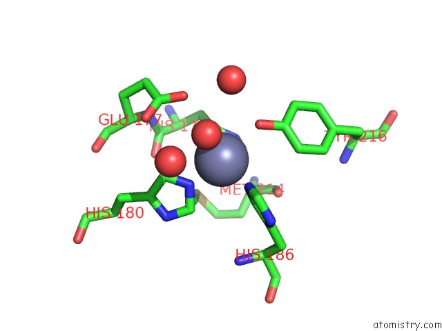
Mono view
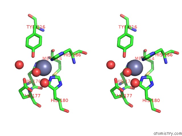
Stereo pair view
Mono view
Stereo pair view
A full contact list of Zinc with other atoms in the Zn binding
site number 1 of Crystal Structure of Serralysin within 5.0Å range:
|
Reference:
D.X.Wu,
T.T.Ran,
W.W.Wang,
D.Q.Xu.
Structure of A Thermostable Serralysin From Serratia Sp. FS14 at 1.1 Angstrom Resolution Acta Crystallogr.,Sect.F V. 72 10 2016.
ISSN: ESSN 2053-230X
DOI: 10.1107/S2053230X15023092
Page generated: Sun Oct 27 14:40:27 2024
ISSN: ESSN 2053-230X
DOI: 10.1107/S2053230X15023092
Last articles
Zn in 9J0NZn in 9J0O
Zn in 9J0P
Zn in 9FJX
Zn in 9EKB
Zn in 9C0F
Zn in 9CAH
Zn in 9CH0
Zn in 9CH3
Zn in 9CH1