Zinc »
PDB 5cdt-5cvm »
5cqk »
Zinc in PDB 5cqk: Crystal Structure of the Cancer Genomic Dna Mutator APOBEC3B
Protein crystallography data
The structure of Crystal Structure of the Cancer Genomic Dna Mutator APOBEC3B, PDB code: 5cqk
was solved by
K.Shi,
K.Kurahashi,
H.Aihara,
with X-Ray Crystallography technique. A brief refinement statistics is given in the table below:
| Resolution Low / High (Å) | 41.92 / 1.88 |
| Space group | P 21 21 21 |
| Cell size a, b, c (Å), α, β, γ (°) | 50.630, 54.040, 66.420, 90.00, 90.00, 90.00 |
| R / Rfree (%) | 17.4 / 20.3 |
Other elements in 5cqk:
The structure of Crystal Structure of the Cancer Genomic Dna Mutator APOBEC3B also contains other interesting chemical elements:
| Sodium | (Na) | 1 atom |
Zinc Binding Sites:
The binding sites of Zinc atom in the Crystal Structure of the Cancer Genomic Dna Mutator APOBEC3B
(pdb code 5cqk). This binding sites where shown within
5.0 Angstroms radius around Zinc atom.
In total only one binding site of Zinc was determined in the Crystal Structure of the Cancer Genomic Dna Mutator APOBEC3B, PDB code: 5cqk:
In total only one binding site of Zinc was determined in the Crystal Structure of the Cancer Genomic Dna Mutator APOBEC3B, PDB code: 5cqk:
Zinc binding site 1 out of 1 in 5cqk
Go back to
Zinc binding site 1 out
of 1 in the Crystal Structure of the Cancer Genomic Dna Mutator APOBEC3B
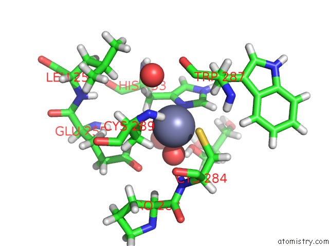
Mono view
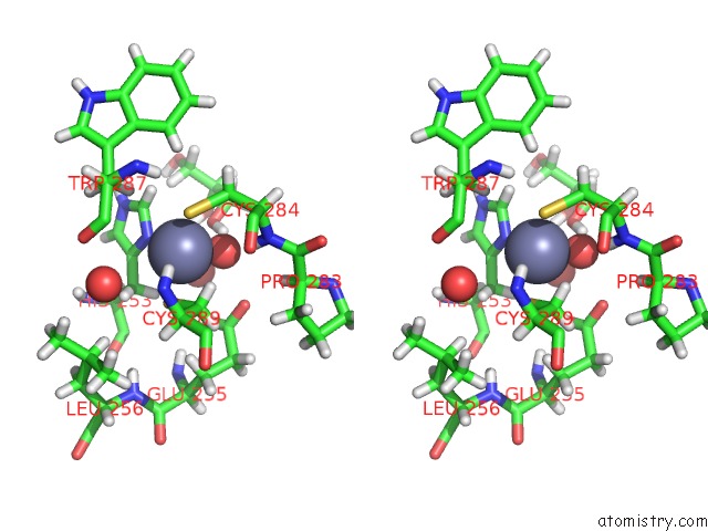
Stereo pair view
Mono view
Stereo pair view
A full contact list of Zinc with other atoms in the Zn binding
site number 1 of Crystal Structure of the Cancer Genomic Dna Mutator APOBEC3B within 5.0Å range:
|
Reference:
K.Shi,
M.A.Carpenter,
K.Kurahashi,
R.S.Harris,
H.Aihara.
Crystal Structure of the Dna Deaminase APOBEC3B Catalytic Domain. J.Biol.Chem. V. 290 28120 2015.
ISSN: ESSN 1083-351X
PubMed: 26416889
DOI: 10.1074/JBC.M115.679951
Page generated: Sun Oct 27 14:27:02 2024
ISSN: ESSN 1083-351X
PubMed: 26416889
DOI: 10.1074/JBC.M115.679951
Last articles
Zn in 9J0NZn in 9J0O
Zn in 9J0P
Zn in 9FJX
Zn in 9EKB
Zn in 9C0F
Zn in 9CAH
Zn in 9CH0
Zn in 9CH3
Zn in 9CH1