Zinc »
PDB 5cdt-5cvm »
5cgx »
Zinc in PDB 5cgx: Crystal Structure of Fox-4 Cephamycinase Mutant Y150F Complexed with Cefoxitin
Enzymatic activity of Crystal Structure of Fox-4 Cephamycinase Mutant Y150F Complexed with Cefoxitin
All present enzymatic activity of Crystal Structure of Fox-4 Cephamycinase Mutant Y150F Complexed with Cefoxitin:
3.5.2.6;
3.5.2.6;
Protein crystallography data
The structure of Crystal Structure of Fox-4 Cephamycinase Mutant Y150F Complexed with Cefoxitin, PDB code: 5cgx
was solved by
V.N.Malashkevich,
R.Toro,
S.Lefurgy,
S.C.Almo,
with X-Ray Crystallography technique. A brief refinement statistics is given in the table below:
| Resolution Low / High (Å) | 19.64 / 1.21 |
| Space group | P 1 21 1 |
| Cell size a, b, c (Å), α, β, γ (°) | 54.524, 56.588, 55.485, 90.00, 99.23, 90.00 |
| R / Rfree (%) | 18.1 / 20.5 |
Other elements in 5cgx:
The structure of Crystal Structure of Fox-4 Cephamycinase Mutant Y150F Complexed with Cefoxitin also contains other interesting chemical elements:
| Sodium | (Na) | 1 atom |
Zinc Binding Sites:
The binding sites of Zinc atom in the Crystal Structure of Fox-4 Cephamycinase Mutant Y150F Complexed with Cefoxitin
(pdb code 5cgx). This binding sites where shown within
5.0 Angstroms radius around Zinc atom.
In total 6 binding sites of Zinc where determined in the Crystal Structure of Fox-4 Cephamycinase Mutant Y150F Complexed with Cefoxitin, PDB code: 5cgx:
Jump to Zinc binding site number: 1; 2; 3; 4; 5; 6;
In total 6 binding sites of Zinc where determined in the Crystal Structure of Fox-4 Cephamycinase Mutant Y150F Complexed with Cefoxitin, PDB code: 5cgx:
Jump to Zinc binding site number: 1; 2; 3; 4; 5; 6;
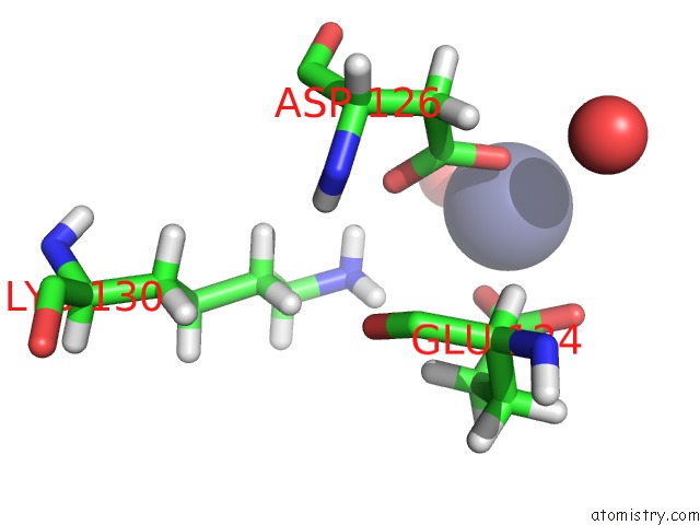
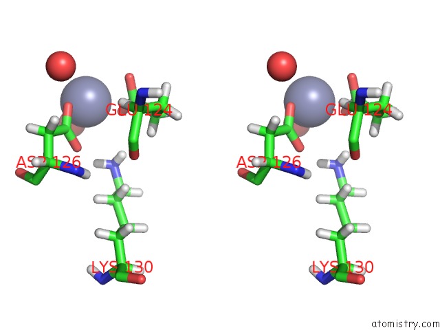
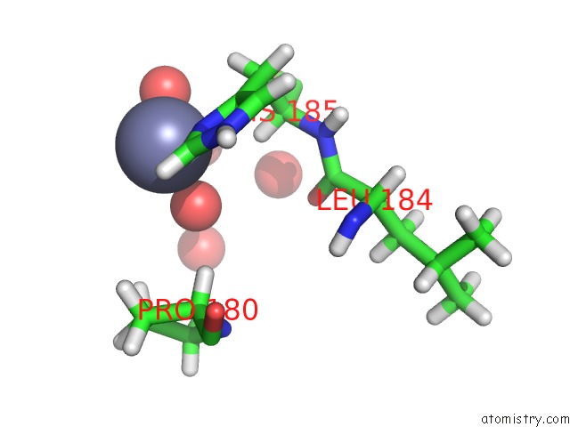
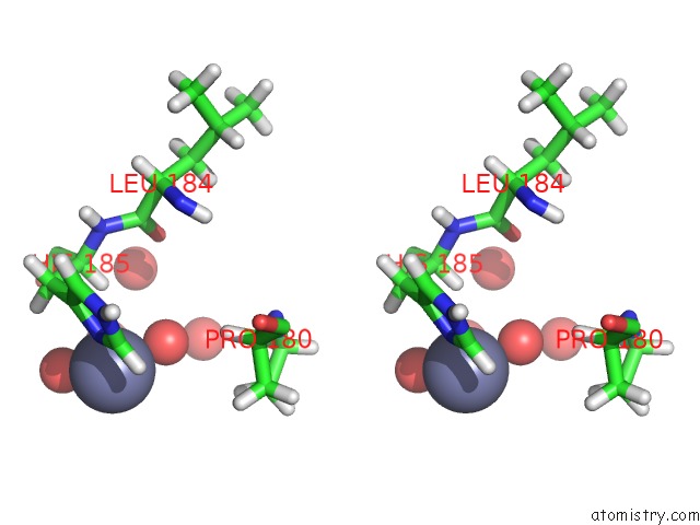
Zinc binding site 1 out of 6 in 5cgx
Go back to
Zinc binding site 1 out
of 6 in the Crystal Structure of Fox-4 Cephamycinase Mutant Y150F Complexed with Cefoxitin
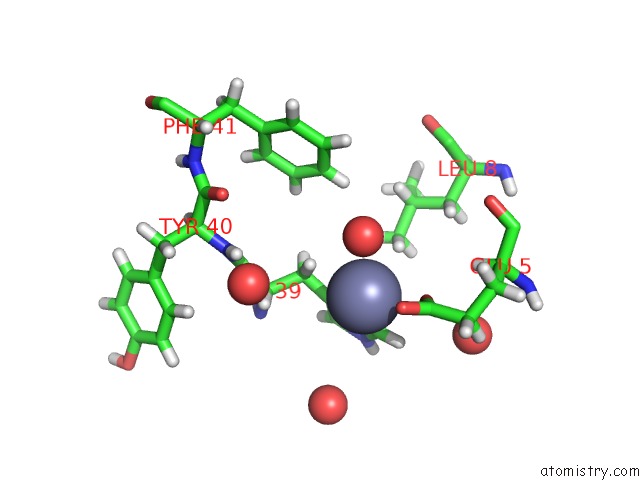
Mono view
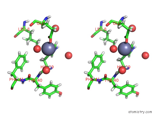
Stereo pair view
Mono view
Stereo pair view
A full contact list of Zinc with other atoms in the Zn binding
site number 1 of Crystal Structure of Fox-4 Cephamycinase Mutant Y150F Complexed with Cefoxitin within 5.0Å range:
|
Zinc binding site 2 out of 6 in 5cgx
Go back to
Zinc binding site 2 out
of 6 in the Crystal Structure of Fox-4 Cephamycinase Mutant Y150F Complexed with Cefoxitin
Mono view
Stereo pair view
Mono view
Stereo pair view
A full contact list of Zinc with other atoms in the Zn binding
site number 2 of Crystal Structure of Fox-4 Cephamycinase Mutant Y150F Complexed with Cefoxitin within 5.0Å range:
|
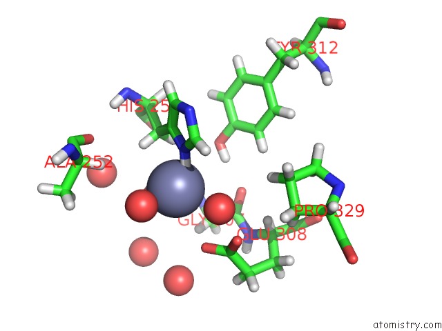
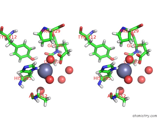
Zinc binding site 3 out of 6 in 5cgx
Go back to
Zinc binding site 3 out
of 6 in the Crystal Structure of Fox-4 Cephamycinase Mutant Y150F Complexed with Cefoxitin
Mono view
Stereo pair view
Mono view
Stereo pair view
A full contact list of Zinc with other atoms in the Zn binding
site number 3 of Crystal Structure of Fox-4 Cephamycinase Mutant Y150F Complexed with Cefoxitin within 5.0Å range:
|
Zinc binding site 4 out of 6 in 5cgx
Go back to
Zinc binding site 4 out
of 6 in the Crystal Structure of Fox-4 Cephamycinase Mutant Y150F Complexed with Cefoxitin
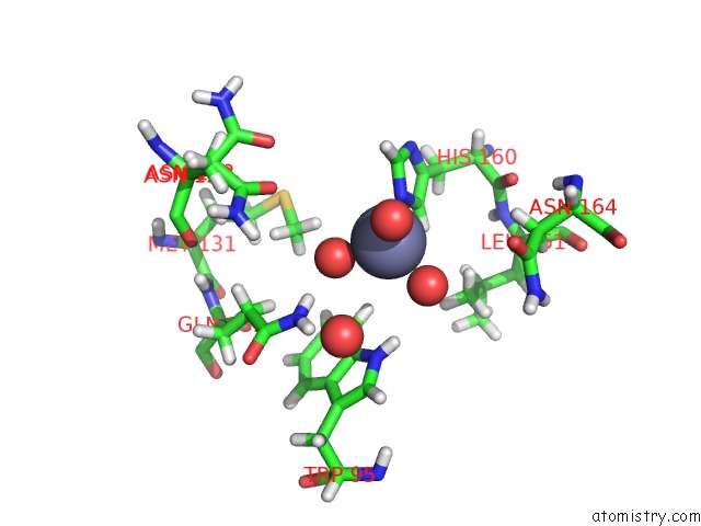
Mono view
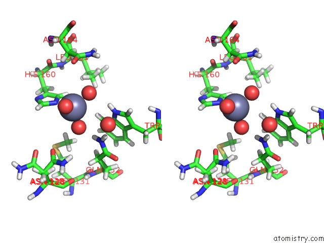
Stereo pair view
Mono view
Stereo pair view
A full contact list of Zinc with other atoms in the Zn binding
site number 4 of Crystal Structure of Fox-4 Cephamycinase Mutant Y150F Complexed with Cefoxitin within 5.0Å range:
|
Zinc binding site 5 out of 6 in 5cgx
Go back to
Zinc binding site 5 out
of 6 in the Crystal Structure of Fox-4 Cephamycinase Mutant Y150F Complexed with Cefoxitin
Mono view
Stereo pair view
Mono view
Stereo pair view
A full contact list of Zinc with other atoms in the Zn binding
site number 5 of Crystal Structure of Fox-4 Cephamycinase Mutant Y150F Complexed with Cefoxitin within 5.0Å range:
|
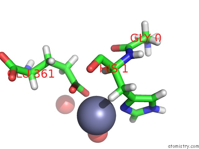
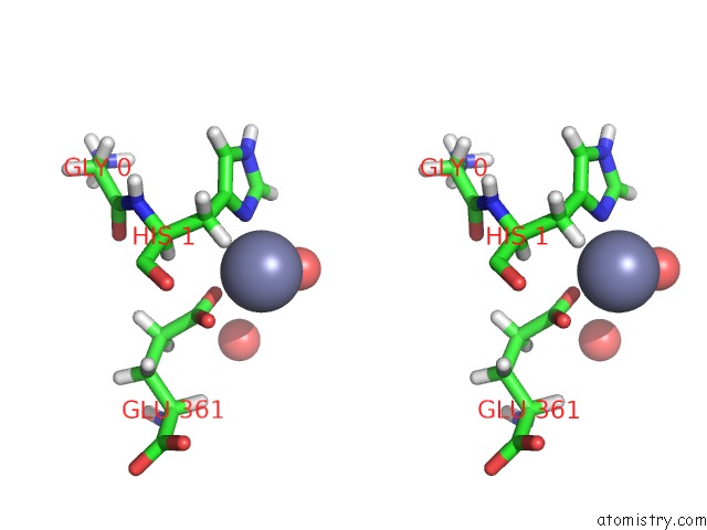
Zinc binding site 6 out of 6 in 5cgx
Go back to
Zinc binding site 6 out
of 6 in the Crystal Structure of Fox-4 Cephamycinase Mutant Y150F Complexed with Cefoxitin
Mono view
Stereo pair view
Mono view
Stereo pair view
A full contact list of Zinc with other atoms in the Zn binding
site number 6 of Crystal Structure of Fox-4 Cephamycinase Mutant Y150F Complexed with Cefoxitin within 5.0Å range:
|
Reference:
S.T.Lefurgy,
V.N.Malashkevich,
J.T.Aguilan,
E.Nieves,
E.C.Mundorff,
B.Biju,
M.A.Noel,
R.Toro,
D.Baiwir,
K.M.Papp-Wallace,
S.C.Almo,
J.M.Frere,
G.Bou,
R.A.Bonomo.
Fox-4 Cephamycinase: An Analysis of Structure and Function. Antimicrob.Agents Chemother. 2015.
ISSN: ESSN 1098-6596
PubMed: 26525784
DOI: 10.1128/AAC.01887-15
Page generated: Sun Oct 27 14:17:41 2024
ISSN: ESSN 1098-6596
PubMed: 26525784
DOI: 10.1128/AAC.01887-15
Last articles
Zn in 9J0NZn in 9J0O
Zn in 9J0P
Zn in 9FJX
Zn in 9EKB
Zn in 9C0F
Zn in 9CAH
Zn in 9CH0
Zn in 9CH3
Zn in 9CH1