Zinc in PDB 5a22: Structure of the L Protein of Vesicular Stomatitis Virus From Electron Cryomicroscopy
Enzymatic activity of Structure of the L Protein of Vesicular Stomatitis Virus From Electron Cryomicroscopy
All present enzymatic activity of Structure of the L Protein of Vesicular Stomatitis Virus From Electron Cryomicroscopy:
2.7.7.6;
2.7.7.6;
Zinc Binding Sites:
The binding sites of Zinc atom in the Structure of the L Protein of Vesicular Stomatitis Virus From Electron Cryomicroscopy
(pdb code 5a22). This binding sites where shown within
5.0 Angstroms radius around Zinc atom.
In total 2 binding sites of Zinc where determined in the Structure of the L Protein of Vesicular Stomatitis Virus From Electron Cryomicroscopy, PDB code: 5a22:
Jump to Zinc binding site number: 1; 2;
In total 2 binding sites of Zinc where determined in the Structure of the L Protein of Vesicular Stomatitis Virus From Electron Cryomicroscopy, PDB code: 5a22:
Jump to Zinc binding site number: 1; 2;
Zinc binding site 1 out of 2 in 5a22
Go back to
Zinc binding site 1 out
of 2 in the Structure of the L Protein of Vesicular Stomatitis Virus From Electron Cryomicroscopy
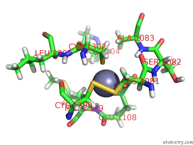
Mono view
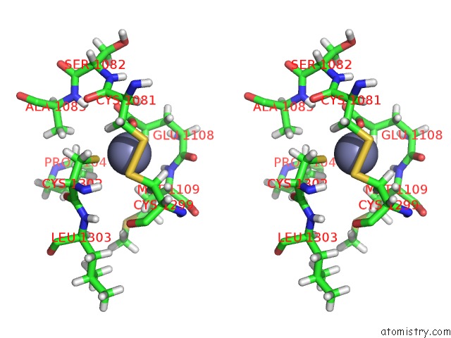
Stereo pair view
Mono view
Stereo pair view
A full contact list of Zinc with other atoms in the Zn binding
site number 1 of Structure of the L Protein of Vesicular Stomatitis Virus From Electron Cryomicroscopy within 5.0Å range:
|
Zinc binding site 2 out of 2 in 5a22
Go back to
Zinc binding site 2 out
of 2 in the Structure of the L Protein of Vesicular Stomatitis Virus From Electron Cryomicroscopy
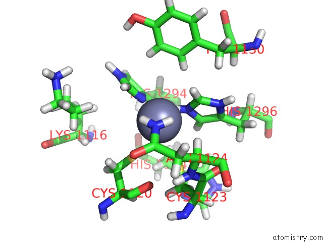
Mono view
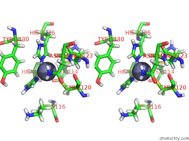
Stereo pair view
Mono view
Stereo pair view
A full contact list of Zinc with other atoms in the Zn binding
site number 2 of Structure of the L Protein of Vesicular Stomatitis Virus From Electron Cryomicroscopy within 5.0Å range:
|
Reference:
B.Liang,
Z.Li,
S.Jenni,
A.A.Rahmeh,
B.M.Morin,
T.Grant,
N.Grigorieff,
S.C.Harrison,
S.P.Whelan.
Structure of the L Protein of Vesicular Stomatitis Virus From Electron Cryomicroscopy. Cell(Cambridge,Mass.) V. 162 314 2015.
ISSN: ISSN 0092-8674
PubMed: 26144317
DOI: 10.1016/J.CELL.2015.06.018
Page generated: Sun Oct 27 12:35:36 2024
ISSN: ISSN 0092-8674
PubMed: 26144317
DOI: 10.1016/J.CELL.2015.06.018
Last articles
Zn in 9MJ5Zn in 9HNW
Zn in 9G0L
Zn in 9FNE
Zn in 9DZN
Zn in 9E0I
Zn in 9D32
Zn in 9DAK
Zn in 8ZXC
Zn in 8ZUF