Zinc »
PDB 4zh3-4zvn »
4zh3 »
Zinc in PDB 4zh3: Crystal Structure of Escherichia Coli Rna Polymerase in Complex with CBRH16-Br
Enzymatic activity of Crystal Structure of Escherichia Coli Rna Polymerase in Complex with CBRH16-Br
All present enzymatic activity of Crystal Structure of Escherichia Coli Rna Polymerase in Complex with CBRH16-Br:
2.7.7.6;
2.7.7.6;
Protein crystallography data
The structure of Crystal Structure of Escherichia Coli Rna Polymerase in Complex with CBRH16-Br, PDB code: 4zh3
was solved by
Y.Feng,
R.H.Ebright,
with X-Ray Crystallography technique. A brief refinement statistics is given in the table below:
| Resolution Low / High (Å) | 49.75 / 4.08 |
| Space group | P 21 21 21 |
| Cell size a, b, c (Å), α, β, γ (°) | 186.339, 205.790, 307.630, 90.00, 90.00, 90.00 |
| R / Rfree (%) | 23.8 / 26.7 |
Other elements in 4zh3:
The structure of Crystal Structure of Escherichia Coli Rna Polymerase in Complex with CBRH16-Br also contains other interesting chemical elements:
| Fluorine | (F) | 8 atoms |
| Magnesium | (Mg) | 2 atoms |
| Bromine | (Br) | 2 atoms |
Zinc Binding Sites:
The binding sites of Zinc atom in the Crystal Structure of Escherichia Coli Rna Polymerase in Complex with CBRH16-Br
(pdb code 4zh3). This binding sites where shown within
5.0 Angstroms radius around Zinc atom.
In total 4 binding sites of Zinc where determined in the Crystal Structure of Escherichia Coli Rna Polymerase in Complex with CBRH16-Br, PDB code: 4zh3:
Jump to Zinc binding site number: 1; 2; 3; 4;
In total 4 binding sites of Zinc where determined in the Crystal Structure of Escherichia Coli Rna Polymerase in Complex with CBRH16-Br, PDB code: 4zh3:
Jump to Zinc binding site number: 1; 2; 3; 4;
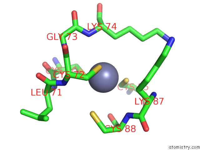
Zinc binding site 1 out of 4 in 4zh3
Go back to
Zinc binding site 1 out
of 4 in the Crystal Structure of Escherichia Coli Rna Polymerase in Complex with CBRH16-Br

Mono view
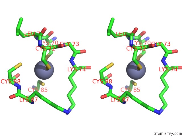
Stereo pair view
Mono view
Stereo pair view
A full contact list of Zinc with other atoms in the Zn binding
site number 1 of Crystal Structure of Escherichia Coli Rna Polymerase in Complex with CBRH16-Br within 5.0Å range:
|
Zinc binding site 2 out of 4 in 4zh3
Go back to
Zinc binding site 2 out
of 4 in the Crystal Structure of Escherichia Coli Rna Polymerase in Complex with CBRH16-Br

Mono view
Stereo pair view
Mono view
Stereo pair view
A full contact list of Zinc with other atoms in the Zn binding
site number 2 of Crystal Structure of Escherichia Coli Rna Polymerase in Complex with CBRH16-Br within 5.0Å range:
|
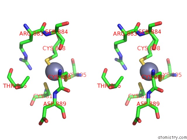
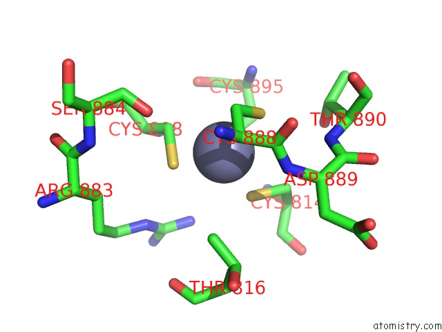
Zinc binding site 3 out of 4 in 4zh3
Go back to
Zinc binding site 3 out
of 4 in the Crystal Structure of Escherichia Coli Rna Polymerase in Complex with CBRH16-Br
Mono view

Stereo pair view
Mono view
Stereo pair view
A full contact list of Zinc with other atoms in the Zn binding
site number 3 of Crystal Structure of Escherichia Coli Rna Polymerase in Complex with CBRH16-Br within 5.0Å range:
|
Zinc binding site 4 out of 4 in 4zh3
Go back to
Zinc binding site 4 out
of 4 in the Crystal Structure of Escherichia Coli Rna Polymerase in Complex with CBRH16-Br
Mono view

Stereo pair view
Mono view
Stereo pair view
A full contact list of Zinc with other atoms in the Zn binding
site number 4 of Crystal Structure of Escherichia Coli Rna Polymerase in Complex with CBRH16-Br within 5.0Å range:
|
Reference:
Y.Feng,
D.Degen,
X.Wang,
M.Gigliotti,
S.Liu,
Y.Zhang,
D.Das,
T.Michalchuk,
Y.W.Ebright,
M.Talaue,
N.Connell,
R.H.Ebright.
Structural Basis of Transcription Inhibition By Cbr Hydroxamidines and Cbr Pyrazoles. Structure V. 23 1470 2015.
ISSN: ISSN 0969-2126
PubMed: 26190576
DOI: 10.1016/J.STR.2015.06.009
Page generated: Sun Oct 27 11:47:57 2024
ISSN: ISSN 0969-2126
PubMed: 26190576
DOI: 10.1016/J.STR.2015.06.009
Last articles
Zn in 9J0NZn in 9J0O
Zn in 9J0P
Zn in 9FJX
Zn in 9EKB
Zn in 9C0F
Zn in 9CAH
Zn in 9CH0
Zn in 9CH3
Zn in 9CH1