Zinc »
PDB 4uyo-4w8y »
4v1z »
Zinc in PDB 4v1z: The 3-D Structure of the Cellobiohydrolase, CEL7A, From Aspergillus Fumigatus
Enzymatic activity of The 3-D Structure of the Cellobiohydrolase, CEL7A, From Aspergillus Fumigatus
All present enzymatic activity of The 3-D Structure of the Cellobiohydrolase, CEL7A, From Aspergillus Fumigatus:
3.2.1.91;
3.2.1.91;
Protein crystallography data
The structure of The 3-D Structure of the Cellobiohydrolase, CEL7A, From Aspergillus Fumigatus, PDB code: 4v1z
was solved by
O.V.Moroz,
M.Maranta,
T.Shaghasi,
P.V.Harris,
K.S.Wilson,
G.J.Davies,
with X-Ray Crystallography technique. A brief refinement statistics is given in the table below:
| Resolution Low / High (Å) | 67.85 / 1.78 |
| Space group | P 21 21 2 |
| Cell size a, b, c (Å), α, β, γ (°) | 79.230, 131.390, 45.920, 90.00, 90.00, 90.00 |
| R / Rfree (%) | 14.406 / 18.895 |
Zinc Binding Sites:
The binding sites of Zinc atom in the The 3-D Structure of the Cellobiohydrolase, CEL7A, From Aspergillus Fumigatus
(pdb code 4v1z). This binding sites where shown within
5.0 Angstroms radius around Zinc atom.
In total 2 binding sites of Zinc where determined in the The 3-D Structure of the Cellobiohydrolase, CEL7A, From Aspergillus Fumigatus, PDB code: 4v1z:
Jump to Zinc binding site number: 1; 2;
In total 2 binding sites of Zinc where determined in the The 3-D Structure of the Cellobiohydrolase, CEL7A, From Aspergillus Fumigatus, PDB code: 4v1z:
Jump to Zinc binding site number: 1; 2;
Zinc binding site 1 out of 2 in 4v1z
Go back to
Zinc binding site 1 out
of 2 in the The 3-D Structure of the Cellobiohydrolase, CEL7A, From Aspergillus Fumigatus
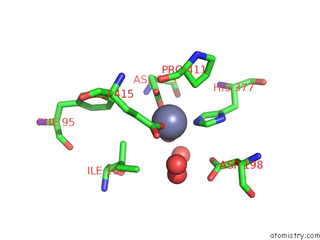
Mono view
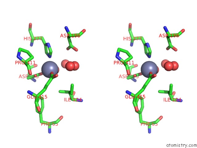
Stereo pair view
Mono view
Stereo pair view
A full contact list of Zinc with other atoms in the Zn binding
site number 1 of The 3-D Structure of the Cellobiohydrolase, CEL7A, From Aspergillus Fumigatus within 5.0Å range:
|
Zinc binding site 2 out of 2 in 4v1z
Go back to
Zinc binding site 2 out
of 2 in the The 3-D Structure of the Cellobiohydrolase, CEL7A, From Aspergillus Fumigatus
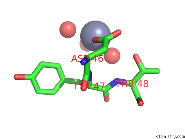
Mono view
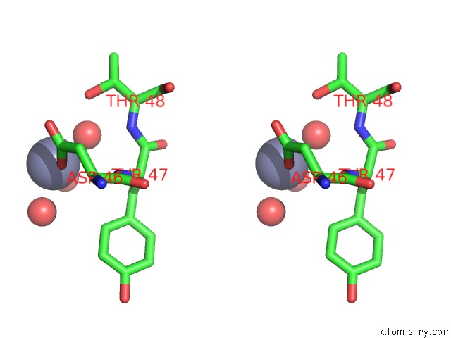
Stereo pair view
Mono view
Stereo pair view
A full contact list of Zinc with other atoms in the Zn binding
site number 2 of The 3-D Structure of the Cellobiohydrolase, CEL7A, From Aspergillus Fumigatus within 5.0Å range:
|
Reference:
O.V.Moroz,
M.Maranta,
T.Shaghasi,
P.V.Harris,
K.S.Wilson,
G.J.Davies.
The Three-Dimensional Structure of the Cellobiohydrolase CEL7A From Aspergillus Fumigatus at 1.5 A Resolution Acta Crystallogr.,Sect.F V. 71 114 2015.
ISSN: ISSN 1744-3091
PubMed: 25615982
DOI: 10.1107/S2053230X14027307
Page generated: Wed Aug 20 22:53:02 2025
ISSN: ISSN 1744-3091
PubMed: 25615982
DOI: 10.1107/S2053230X14027307
Last articles
Zn in 5LSVZn in 5LSZ
Zn in 5LSY
Zn in 5LSU
Zn in 5LSX
Zn in 5LSS
Zn in 5LSC
Zn in 5LST
Zn in 5LPI
Zn in 5LS3