Zinc in PDB 4u5g: Crystal Structure of Con-Ikot-Ikot Toxin
Protein crystallography data
The structure of Crystal Structure of Con-Ikot-Ikot Toxin, PDB code: 4u5g
was solved by
L.Chen,
E.Gouaux,
with X-Ray Crystallography technique. A brief refinement statistics is given in the table below:
| Resolution Low / High (Å) | 19.83 / 2.20 |
| Space group | P 31 2 1 |
| Cell size a, b, c (Å), α, β, γ (°) | 56.499, 56.499, 86.782, 90.00, 90.00, 120.00 |
| R / Rfree (%) | 20.2 / 23.9 |
Zinc Binding Sites:
The binding sites of Zinc atom in the Crystal Structure of Con-Ikot-Ikot Toxin
(pdb code 4u5g). This binding sites where shown within
5.0 Angstroms radius around Zinc atom.
In total 2 binding sites of Zinc where determined in the Crystal Structure of Con-Ikot-Ikot Toxin, PDB code: 4u5g:
Jump to Zinc binding site number: 1; 2;
In total 2 binding sites of Zinc where determined in the Crystal Structure of Con-Ikot-Ikot Toxin, PDB code: 4u5g:
Jump to Zinc binding site number: 1; 2;
Zinc binding site 1 out of 2 in 4u5g
Go back to
Zinc binding site 1 out
of 2 in the Crystal Structure of Con-Ikot-Ikot Toxin
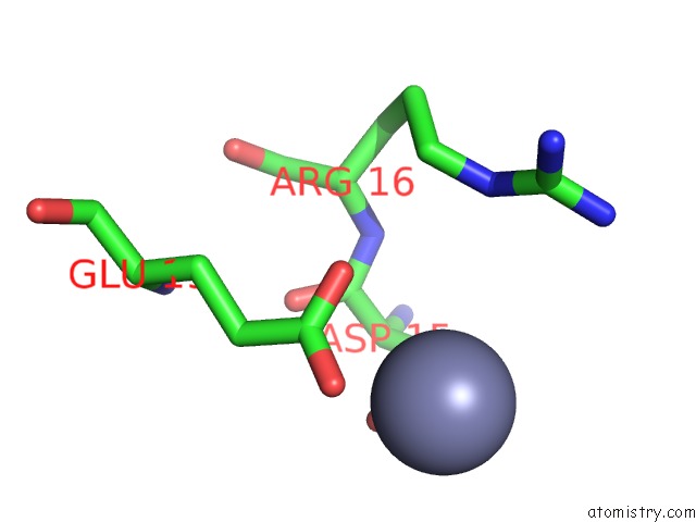
Mono view
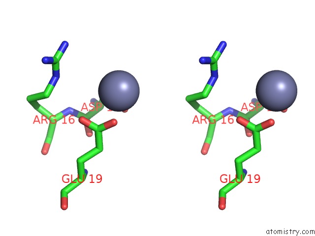
Stereo pair view
Mono view
Stereo pair view
A full contact list of Zinc with other atoms in the Zn binding
site number 1 of Crystal Structure of Con-Ikot-Ikot Toxin within 5.0Å range:
|
Zinc binding site 2 out of 2 in 4u5g
Go back to
Zinc binding site 2 out
of 2 in the Crystal Structure of Con-Ikot-Ikot Toxin
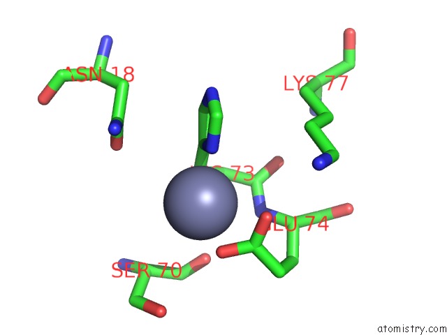
Mono view
Stereo pair view
Mono view
Stereo pair view
A full contact list of Zinc with other atoms in the Zn binding
site number 2 of Crystal Structure of Con-Ikot-Ikot Toxin within 5.0Å range:
|
Reference:
L.Chen,
K.L.Durr,
E.Gouaux.
X-Ray Structures of Ampa Receptor-Cone Snail Toxin Complexes Illuminate Activation Mechanism. Science V. 345 1021 2014.
ISSN: ESSN 1095-9203
PubMed: 25103405
DOI: 10.1126/SCIENCE.1258409
Page generated: Sun Oct 27 08:53:00 2024
ISSN: ESSN 1095-9203
PubMed: 25103405
DOI: 10.1126/SCIENCE.1258409
Last articles
Zn in 9MJ5Zn in 9HNW
Zn in 9G0L
Zn in 9FNE
Zn in 9DZN
Zn in 9E0I
Zn in 9D32
Zn in 9DAK
Zn in 8ZXC
Zn in 8ZUF