Zinc »
PDB 4o8x-4ojv »
4oew »
Zinc in PDB 4oew: Crystal Structure of the PDE5A1 Catalytic Domain in Complex with Novel Inhibitors
Enzymatic activity of Crystal Structure of the PDE5A1 Catalytic Domain in Complex with Novel Inhibitors
All present enzymatic activity of Crystal Structure of the PDE5A1 Catalytic Domain in Complex with Novel Inhibitors:
3.1.4.35;
3.1.4.35;
Protein crystallography data
The structure of Crystal Structure of the PDE5A1 Catalytic Domain in Complex with Novel Inhibitors, PDB code: 4oew
was solved by
T.T.Chen,
J.Ren,
Y.C.Xu,
with X-Ray Crystallography technique. A brief refinement statistics is given in the table below:
| Resolution Low / High (Å) | 32.09 / 2.44 |
| Space group | P 31 2 1 |
| Cell size a, b, c (Å), α, β, γ (°) | 74.120, 74.120, 131.800, 90.00, 90.00, 120.00 |
| R / Rfree (%) | 20.6 / 25.9 |
Other elements in 4oew:
The structure of Crystal Structure of the PDE5A1 Catalytic Domain in Complex with Novel Inhibitors also contains other interesting chemical elements:
| Magnesium | (Mg) | 1 atom |
| Iodine | (I) | 1 atom |
Zinc Binding Sites:
The binding sites of Zinc atom in the Crystal Structure of the PDE5A1 Catalytic Domain in Complex with Novel Inhibitors
(pdb code 4oew). This binding sites where shown within
5.0 Angstroms radius around Zinc atom.
In total only one binding site of Zinc was determined in the Crystal Structure of the PDE5A1 Catalytic Domain in Complex with Novel Inhibitors, PDB code: 4oew:
In total only one binding site of Zinc was determined in the Crystal Structure of the PDE5A1 Catalytic Domain in Complex with Novel Inhibitors, PDB code: 4oew:
Zinc binding site 1 out of 1 in 4oew
Go back to
Zinc binding site 1 out
of 1 in the Crystal Structure of the PDE5A1 Catalytic Domain in Complex with Novel Inhibitors
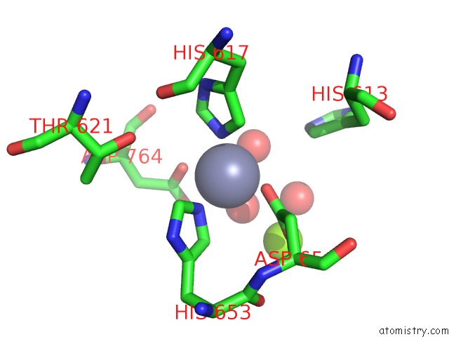
Mono view
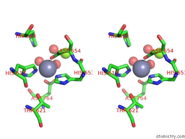
Stereo pair view
Mono view
Stereo pair view
A full contact list of Zinc with other atoms in the Zn binding
site number 1 of Crystal Structure of the PDE5A1 Catalytic Domain in Complex with Novel Inhibitors within 5.0Å range:
|
Reference:
J.Ren,
Y.He,
W.Chen,
T.Chen,
G.Wang,
Z.Wang,
Z.Xu,
X.Luo,
W.Zhu,
H.Jiang,
J.Shen,
Y.Xu.
Thermodynamic and Structural Characterization of Halogen Bonding in Protein-Ligand Interactions: A Case Study of PDE5 and Its Inhibitors. J.Med.Chem. V. 57 3588 2014.
ISSN: ISSN 0022-2623
PubMed: 24702184
DOI: 10.1021/JM5002315
Page generated: Sun Oct 27 03:41:01 2024
ISSN: ISSN 0022-2623
PubMed: 24702184
DOI: 10.1021/JM5002315
Last articles
Zn in 9J0NZn in 9J0O
Zn in 9J0P
Zn in 9FJX
Zn in 9EKB
Zn in 9C0F
Zn in 9CAH
Zn in 9CH0
Zn in 9CH3
Zn in 9CH1