Zinc »
PDB 4o8x-4ojv »
4ocm »
Zinc in PDB 4ocm: Crystal Structure of the RPN8-RPN11 Mpn Domain Heterodimer, Crystal Form Ib
Protein crystallography data
The structure of Crystal Structure of the RPN8-RPN11 Mpn Domain Heterodimer, Crystal Form Ib, PDB code: 4ocm
was solved by
G.R.Pathare,
A.Bracher,
with X-Ray Crystallography technique. A brief refinement statistics is given in the table below:
| Resolution Low / High (Å) | 30.00 / 1.99 |
| Space group | P 1 2 1 |
| Cell size a, b, c (Å), α, β, γ (°) | 63.401, 44.970, 200.043, 90.00, 98.40, 90.00 |
| R / Rfree (%) | 21.6 / 26.2 |
Other elements in 4ocm:
The structure of Crystal Structure of the RPN8-RPN11 Mpn Domain Heterodimer, Crystal Form Ib also contains other interesting chemical elements:
| Potassium | (K) | 1 atom |
Zinc Binding Sites:
The binding sites of Zinc atom in the Crystal Structure of the RPN8-RPN11 Mpn Domain Heterodimer, Crystal Form Ib
(pdb code 4ocm). This binding sites where shown within
5.0 Angstroms radius around Zinc atom.
In total 2 binding sites of Zinc where determined in the Crystal Structure of the RPN8-RPN11 Mpn Domain Heterodimer, Crystal Form Ib, PDB code: 4ocm:
Jump to Zinc binding site number: 1; 2;
In total 2 binding sites of Zinc where determined in the Crystal Structure of the RPN8-RPN11 Mpn Domain Heterodimer, Crystal Form Ib, PDB code: 4ocm:
Jump to Zinc binding site number: 1; 2;
Zinc binding site 1 out of 2 in 4ocm
Go back to
Zinc binding site 1 out
of 2 in the Crystal Structure of the RPN8-RPN11 Mpn Domain Heterodimer, Crystal Form Ib

Mono view
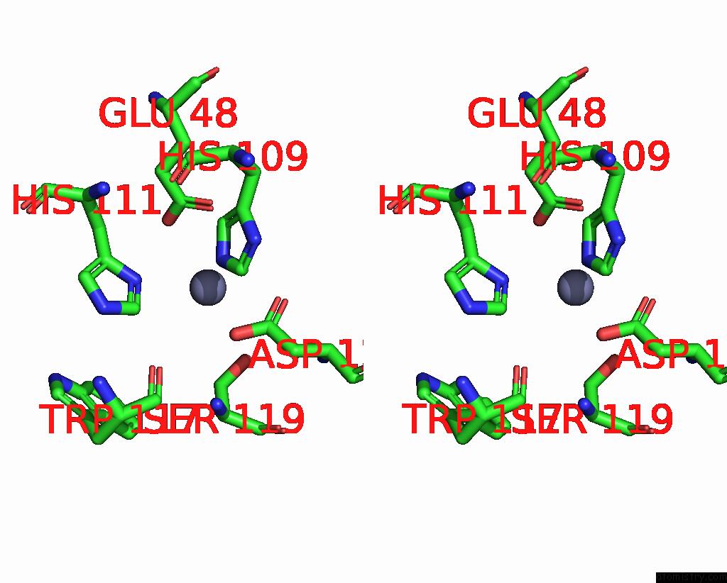
Stereo pair view
Mono view
Stereo pair view
A full contact list of Zinc with other atoms in the Zn binding
site number 1 of Crystal Structure of the RPN8-RPN11 Mpn Domain Heterodimer, Crystal Form Ib within 5.0Å range:
|
Zinc binding site 2 out of 2 in 4ocm
Go back to
Zinc binding site 2 out
of 2 in the Crystal Structure of the RPN8-RPN11 Mpn Domain Heterodimer, Crystal Form Ib
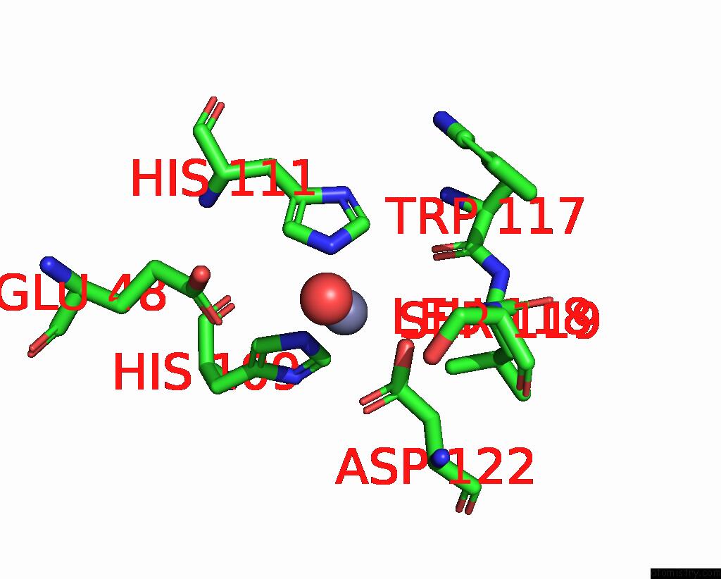
Mono view
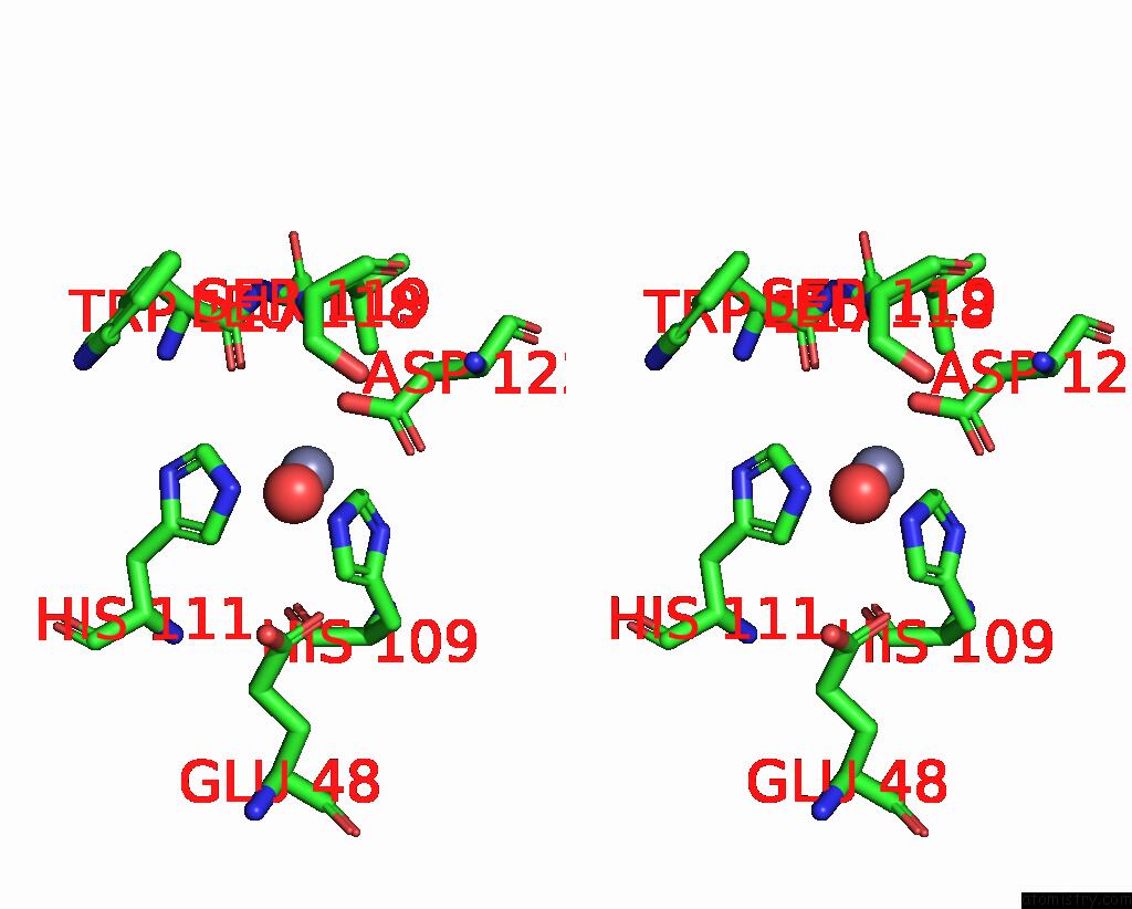
Stereo pair view
Mono view
Stereo pair view
A full contact list of Zinc with other atoms in the Zn binding
site number 2 of Crystal Structure of the RPN8-RPN11 Mpn Domain Heterodimer, Crystal Form Ib within 5.0Å range:
|
Reference:
G.R.Pathare,
I.Nagy,
P.Sledz,
D.J.Anderson,
H.J.Zhou,
E.Pardon,
J.Steyaert,
F.Forster,
A.Bracher,
W.Baumeister.
Crystal Structure of the Proteasomal Deubiquitylation Module RPN8-RPN11. Proc.Natl.Acad.Sci.Usa V. 111 2984 2014.
ISSN: ISSN 0027-8424
PubMed: 24516147
DOI: 10.1073/PNAS.1400546111
Page generated: Sun Oct 27 03:40:01 2024
ISSN: ISSN 0027-8424
PubMed: 24516147
DOI: 10.1073/PNAS.1400546111
Last articles
Zn in 9J0NZn in 9J0O
Zn in 9J0P
Zn in 9FJX
Zn in 9EKB
Zn in 9C0F
Zn in 9CAH
Zn in 9CH0
Zn in 9CH3
Zn in 9CH1