Zinc »
PDB 4o8x-4ojv »
4oc3 »
Zinc in PDB 4oc3: X-Ray Structure of of Human Glutamate Carboxypeptidase II (Gcpii) in A Complex with Cfibzl, A Urea-Based Inhibitor N~2~-{[(1S)-1-Carboxy-2- (Furan-2-Yl)Ethyl]Carbamoyl}-N~6~-(4-Iodobenzoyl)-L-Lysine
Enzymatic activity of X-Ray Structure of of Human Glutamate Carboxypeptidase II (Gcpii) in A Complex with Cfibzl, A Urea-Based Inhibitor N~2~-{[(1S)-1-Carboxy-2- (Furan-2-Yl)Ethyl]Carbamoyl}-N~6~-(4-Iodobenzoyl)-L-Lysine
All present enzymatic activity of X-Ray Structure of of Human Glutamate Carboxypeptidase II (Gcpii) in A Complex with Cfibzl, A Urea-Based Inhibitor N~2~-{[(1S)-1-Carboxy-2- (Furan-2-Yl)Ethyl]Carbamoyl}-N~6~-(4-Iodobenzoyl)-L-Lysine:
3.4.17.21;
3.4.17.21;
Protein crystallography data
The structure of X-Ray Structure of of Human Glutamate Carboxypeptidase II (Gcpii) in A Complex with Cfibzl, A Urea-Based Inhibitor N~2~-{[(1S)-1-Carboxy-2- (Furan-2-Yl)Ethyl]Carbamoyl}-N~6~-(4-Iodobenzoyl)-L-Lysine, PDB code: 4oc3
was solved by
J.Pavlicek,
J.Ptacek,
J.Cerny,
Y.Byun,
L.Skultetyova,
M.Pomper,
J.Lubkowski,
C.Barinka,
with X-Ray Crystallography technique. A brief refinement statistics is given in the table below:
| Resolution Low / High (Å) | 29.52 / 1.79 |
| Space group | I 2 2 2 |
| Cell size a, b, c (Å), α, β, γ (°) | 101.566, 130.434, 159.017, 90.00, 90.00, 90.00 |
| R / Rfree (%) | 16.1 / 18.1 |
Other elements in 4oc3:
The structure of X-Ray Structure of of Human Glutamate Carboxypeptidase II (Gcpii) in A Complex with Cfibzl, A Urea-Based Inhibitor N~2~-{[(1S)-1-Carboxy-2- (Furan-2-Yl)Ethyl]Carbamoyl}-N~6~-(4-Iodobenzoyl)-L-Lysine also contains other interesting chemical elements:
| Iodine | (I) | 1 atom |
| Chlorine | (Cl) | 1 atom |
| Calcium | (Ca) | 1 atom |
Zinc Binding Sites:
The binding sites of Zinc atom in the X-Ray Structure of of Human Glutamate Carboxypeptidase II (Gcpii) in A Complex with Cfibzl, A Urea-Based Inhibitor N~2~-{[(1S)-1-Carboxy-2- (Furan-2-Yl)Ethyl]Carbamoyl}-N~6~-(4-Iodobenzoyl)-L-Lysine
(pdb code 4oc3). This binding sites where shown within
5.0 Angstroms radius around Zinc atom.
In total 2 binding sites of Zinc where determined in the X-Ray Structure of of Human Glutamate Carboxypeptidase II (Gcpii) in A Complex with Cfibzl, A Urea-Based Inhibitor N~2~-{[(1S)-1-Carboxy-2- (Furan-2-Yl)Ethyl]Carbamoyl}-N~6~-(4-Iodobenzoyl)-L-Lysine, PDB code: 4oc3:
Jump to Zinc binding site number: 1; 2;
Zinc binding site 1 out of 2 in 4oc3
Go back to
Zinc binding site 1 out
of 2 in the X-Ray Structure of of Human Glutamate Carboxypeptidase II (Gcpii) in A Complex with Cfibzl, A Urea-Based Inhibitor N~2~-{[(1S)-1-Carboxy-2- (Furan-2-Yl)Ethyl]Carbamoyl}-N~6~-(4-Iodobenzoyl)-L-Lysine
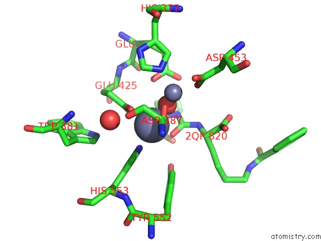
Mono view
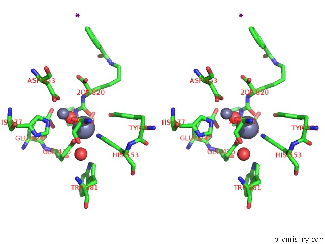
Stereo pair view
Mono view
Stereo pair view
A full contact list of Zinc with other atoms in the Zn binding
site number 1 of X-Ray Structure of of Human Glutamate Carboxypeptidase II (Gcpii) in A Complex with Cfibzl, A Urea-Based Inhibitor N~2~-{[(1S)-1-Carboxy-2- (Furan-2-Yl)Ethyl]Carbamoyl}-N~6~-(4-Iodobenzoyl)-L-Lysine within 5.0Å range:
|
Zinc binding site 2 out of 2 in 4oc3
Go back to
Zinc binding site 2 out
of 2 in the X-Ray Structure of of Human Glutamate Carboxypeptidase II (Gcpii) in A Complex with Cfibzl, A Urea-Based Inhibitor N~2~-{[(1S)-1-Carboxy-2- (Furan-2-Yl)Ethyl]Carbamoyl}-N~6~-(4-Iodobenzoyl)-L-Lysine
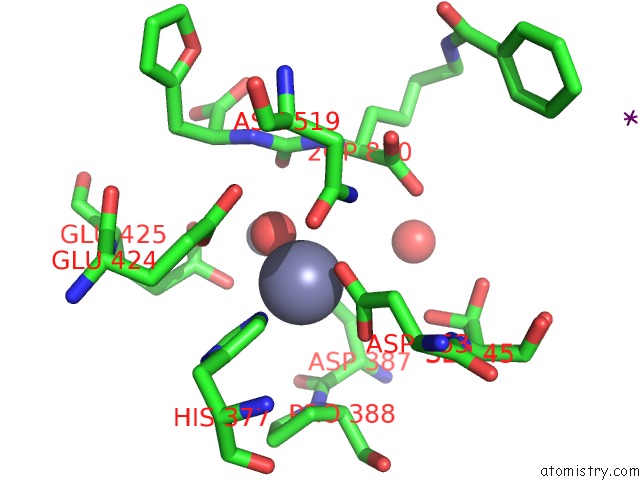
Mono view
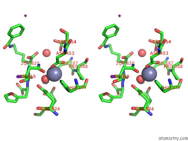
Stereo pair view
Mono view
Stereo pair view
A full contact list of Zinc with other atoms in the Zn binding
site number 2 of X-Ray Structure of of Human Glutamate Carboxypeptidase II (Gcpii) in A Complex with Cfibzl, A Urea-Based Inhibitor N~2~-{[(1S)-1-Carboxy-2- (Furan-2-Yl)Ethyl]Carbamoyl}-N~6~-(4-Iodobenzoyl)-L-Lysine within 5.0Å range:
|
Reference:
J.Pavlicek,
J.Ptacek,
J.Cerny,
Y.Byun,
L.Skultetyova,
M.G.Pomper,
J.Lubkowski,
C.Barinka.
Structural Characterization of P1'-Diversified Urea-Based Inhibitors of Glutamate Carboxypeptidase II. Bioorg.Med.Chem.Lett. V. 24 2340 2014.
ISSN: ISSN 0960-894X
PubMed: 24731280
DOI: 10.1016/J.BMCL.2014.03.066
Page generated: Sun Oct 27 03:38:56 2024
ISSN: ISSN 0960-894X
PubMed: 24731280
DOI: 10.1016/J.BMCL.2014.03.066
Last articles
Zn in 9J0NZn in 9J0O
Zn in 9J0P
Zn in 9FJX
Zn in 9EKB
Zn in 9C0F
Zn in 9CAH
Zn in 9CH0
Zn in 9CH3
Zn in 9CH1