Zinc »
PDB 4ngs-4nu7 »
4niy »
Zinc in PDB 4niy: Crystal Structure of Trypsiligase (K60E/N143H/Y151H/D189K Trypsin) Complexed to Yrh-Ecotin (M84Y/M85R/A86H Ecotin)
Enzymatic activity of Crystal Structure of Trypsiligase (K60E/N143H/Y151H/D189K Trypsin) Complexed to Yrh-Ecotin (M84Y/M85R/A86H Ecotin)
All present enzymatic activity of Crystal Structure of Trypsiligase (K60E/N143H/Y151H/D189K Trypsin) Complexed to Yrh-Ecotin (M84Y/M85R/A86H Ecotin):
3.4.21.4;
3.4.21.4;
Protein crystallography data
The structure of Crystal Structure of Trypsiligase (K60E/N143H/Y151H/D189K Trypsin) Complexed to Yrh-Ecotin (M84Y/M85R/A86H Ecotin), PDB code: 4niy
was solved by
M.Schoepfel,
C.Parthier,
M.T.Stubbs,
with X-Ray Crystallography technique. A brief refinement statistics is given in the table below:
| Resolution Low / High (Å) | 49.60 / 2.84 |
| Space group | P 1 21 1 |
| Cell size a, b, c (Å), α, β, γ (°) | 94.241, 78.615, 98.084, 90.00, 96.62, 90.00 |
| R / Rfree (%) | 19.2 / 25.2 |
Other elements in 4niy:
The structure of Crystal Structure of Trypsiligase (K60E/N143H/Y151H/D189K Trypsin) Complexed to Yrh-Ecotin (M84Y/M85R/A86H Ecotin) also contains other interesting chemical elements:
| Calcium | (Ca) | 4 atoms |
Zinc Binding Sites:
The binding sites of Zinc atom in the Crystal Structure of Trypsiligase (K60E/N143H/Y151H/D189K Trypsin) Complexed to Yrh-Ecotin (M84Y/M85R/A86H Ecotin)
(pdb code 4niy). This binding sites where shown within
5.0 Angstroms radius around Zinc atom.
In total only one binding site of Zinc was determined in the Crystal Structure of Trypsiligase (K60E/N143H/Y151H/D189K Trypsin) Complexed to Yrh-Ecotin (M84Y/M85R/A86H Ecotin), PDB code: 4niy:
In total only one binding site of Zinc was determined in the Crystal Structure of Trypsiligase (K60E/N143H/Y151H/D189K Trypsin) Complexed to Yrh-Ecotin (M84Y/M85R/A86H Ecotin), PDB code: 4niy:
Zinc binding site 1 out of 1 in 4niy
Go back to
Zinc binding site 1 out
of 1 in the Crystal Structure of Trypsiligase (K60E/N143H/Y151H/D189K Trypsin) Complexed to Yrh-Ecotin (M84Y/M85R/A86H Ecotin)
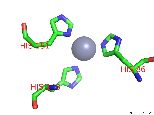
Mono view
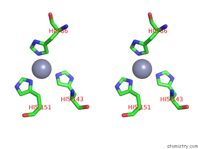
Stereo pair view
Mono view
Stereo pair view
A full contact list of Zinc with other atoms in the Zn binding
site number 1 of Crystal Structure of Trypsiligase (K60E/N143H/Y151H/D189K Trypsin) Complexed to Yrh-Ecotin (M84Y/M85R/A86H Ecotin) within 5.0Å range:
|
Reference:
S.Liebscher,
M.Schopfel,
T.Aumuller,
A.Sharkhuukhen,
A.Pech,
E.Hoss,
C.Parthier,
G.Jahreis,
M.T.Stubbs,
F.Bordusa.
N-Terminal Protein Modification By Substrate-Activated Reverse Proteolysis. Angew.Chem.Int.Ed.Engl. V. 53 3024 2014.
ISSN: ISSN 1433-7851
PubMed: 24520050
DOI: 10.1002/ANIE.201307736
Page generated: Sun Oct 27 03:11:58 2024
ISSN: ISSN 1433-7851
PubMed: 24520050
DOI: 10.1002/ANIE.201307736
Last articles
Zn in 9J0NZn in 9J0O
Zn in 9J0P
Zn in 9FJX
Zn in 9EKB
Zn in 9C0F
Zn in 9CAH
Zn in 9CH0
Zn in 9CH3
Zn in 9CH1