Zinc »
PDB 4n4e-4ngr »
4nef »
Zinc in PDB 4nef: X-Ray Structure of Human Aquaporin 2
Protein crystallography data
The structure of X-Ray Structure of Human Aquaporin 2, PDB code: 4nef
was solved by
A.Frick,
U.Eriksson,
F.D.Mattia,
F.Oberg,
K.Hedfalk,
R.Neutze,
W.D.Grip,
P.M.T.Deen,
S.Tornroth-Horsefield,
with X-Ray Crystallography technique. A brief refinement statistics is given in the table below:
| Resolution Low / High (Å) | 49.96 / 2.75 |
| Space group | P 42 |
| Cell size a, b, c (Å), α, β, γ (°) | 119.110, 119.110, 90.621, 90.00, 90.00, 90.00 |
| R / Rfree (%) | 20.2 / 22.5 |
Other elements in 4nef:
The structure of X-Ray Structure of Human Aquaporin 2 also contains other interesting chemical elements:
| Cadmium | (Cd) | 2 atoms |
Zinc Binding Sites:
The binding sites of Zinc atom in the X-Ray Structure of Human Aquaporin 2
(pdb code 4nef). This binding sites where shown within
5.0 Angstroms radius around Zinc atom.
In total only one binding site of Zinc was determined in the X-Ray Structure of Human Aquaporin 2, PDB code: 4nef:
In total only one binding site of Zinc was determined in the X-Ray Structure of Human Aquaporin 2, PDB code: 4nef:
Zinc binding site 1 out of 1 in 4nef
Go back to
Zinc binding site 1 out
of 1 in the X-Ray Structure of Human Aquaporin 2
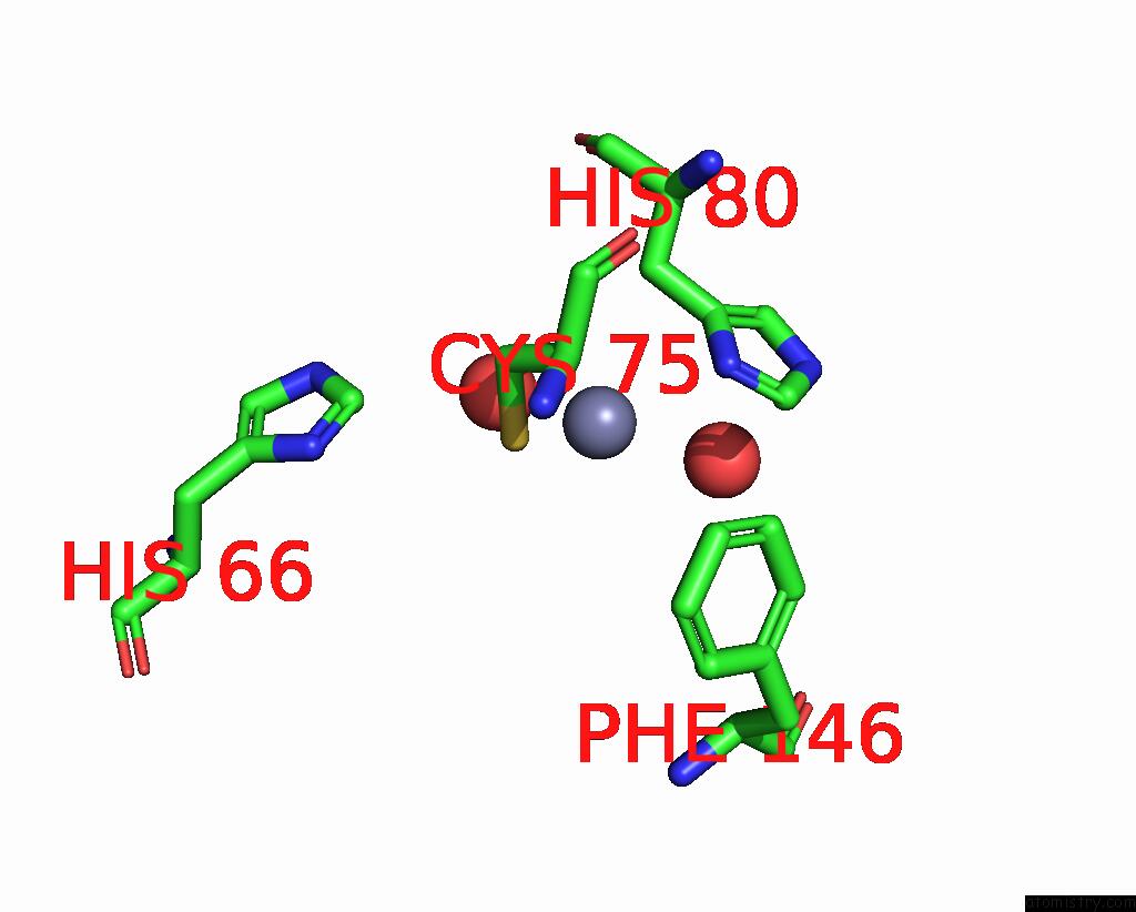
Mono view
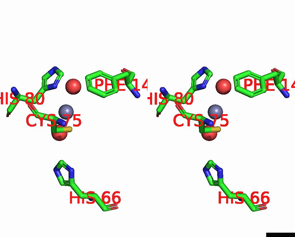
Stereo pair view
Mono view
Stereo pair view
A full contact list of Zinc with other atoms in the Zn binding
site number 1 of X-Ray Structure of Human Aquaporin 2 within 5.0Å range:
|
Reference:
A.Frick,
U.Eriksson,
F.D.Mattia,
F.Oberg,
K.Hedfalk,
R.Neutze,
W.D.Grip,
P.M.T.Deen,
S.Tornroth-Horsefield.
X-Ray Structure of Human Aquapor in 2 and Its Implications For Nephrogenic Diabetes Insipidus and Trafficking Proc.Natl.Acad.Sci.Usa 2014.
ISSN: ESSN 1091-6490
Page generated: Sun Oct 27 03:06:15 2024
ISSN: ESSN 1091-6490
Last articles
Zn in 9J0NZn in 9J0O
Zn in 9J0P
Zn in 9FJX
Zn in 9EKB
Zn in 9C0F
Zn in 9CAH
Zn in 9CH0
Zn in 9CH3
Zn in 9CH1