Zinc »
PDB 4m3p-4mi0 »
4mdt »
Zinc in PDB 4mdt: Structure of Lpxc Bound to the Reaction Product Udp-(3-O-(R-3- Hydroxymyristoyl))-Glucosamine
Protein crystallography data
The structure of Structure of Lpxc Bound to the Reaction Product Udp-(3-O-(R-3- Hydroxymyristoyl))-Glucosamine, PDB code: 4mdt
was solved by
G.M.Clayton,
D.J.Klein,
K.W.Rickert,
S.B.Patel,
M.Kornienko,
J.Zugay-Murphy,
J.C.Reid,
S.Tummala,
S.Sharma,
S.B.Singh,
L.Miesel,
K.J.Lumb,
S.M.Soisson,
with X-Ray Crystallography technique. A brief refinement statistics is given in the table below:
| Resolution Low / High (Å) | 50.45 / 2.59 |
| Space group | C 1 2 1 |
| Cell size a, b, c (Å), α, β, γ (°) | 168.973, 103.520, 103.968, 90.00, 103.96, 90.00 |
| R / Rfree (%) | 19.7 / 24 |
Zinc Binding Sites:
The binding sites of Zinc atom in the Structure of Lpxc Bound to the Reaction Product Udp-(3-O-(R-3- Hydroxymyristoyl))-Glucosamine
(pdb code 4mdt). This binding sites where shown within
5.0 Angstroms radius around Zinc atom.
In total 4 binding sites of Zinc where determined in the Structure of Lpxc Bound to the Reaction Product Udp-(3-O-(R-3- Hydroxymyristoyl))-Glucosamine, PDB code: 4mdt:
Jump to Zinc binding site number: 1; 2; 3; 4;
In total 4 binding sites of Zinc where determined in the Structure of Lpxc Bound to the Reaction Product Udp-(3-O-(R-3- Hydroxymyristoyl))-Glucosamine, PDB code: 4mdt:
Jump to Zinc binding site number: 1; 2; 3; 4;
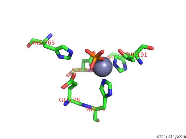
Zinc binding site 1 out of 4 in 4mdt
Go back to
Zinc binding site 1 out
of 4 in the Structure of Lpxc Bound to the Reaction Product Udp-(3-O-(R-3- Hydroxymyristoyl))-Glucosamine
Mono view
Stereo pair view
Mono view
Stereo pair view
A full contact list of Zinc with other atoms in the Zn binding
site number 1 of Structure of Lpxc Bound to the Reaction Product Udp-(3-O-(R-3- Hydroxymyristoyl))-Glucosamine within 5.0Å range:
|
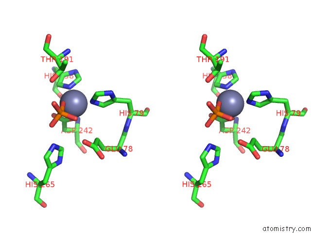
Zinc binding site 2 out of 4 in 4mdt
Go back to
Zinc binding site 2 out
of 4 in the Structure of Lpxc Bound to the Reaction Product Udp-(3-O-(R-3- Hydroxymyristoyl))-Glucosamine
Mono view

Stereo pair view
Mono view
Stereo pair view
A full contact list of Zinc with other atoms in the Zn binding
site number 2 of Structure of Lpxc Bound to the Reaction Product Udp-(3-O-(R-3- Hydroxymyristoyl))-Glucosamine within 5.0Å range:
|
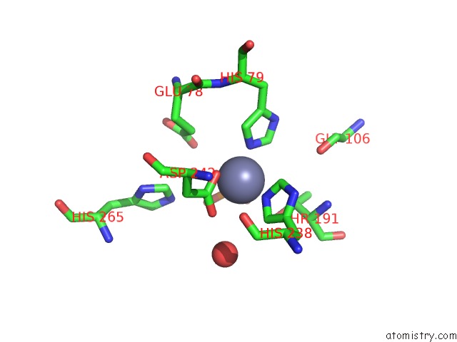
Zinc binding site 3 out of 4 in 4mdt
Go back to
Zinc binding site 3 out
of 4 in the Structure of Lpxc Bound to the Reaction Product Udp-(3-O-(R-3- Hydroxymyristoyl))-Glucosamine
Mono view

Stereo pair view
Mono view
Stereo pair view
A full contact list of Zinc with other atoms in the Zn binding
site number 3 of Structure of Lpxc Bound to the Reaction Product Udp-(3-O-(R-3- Hydroxymyristoyl))-Glucosamine within 5.0Å range:
|
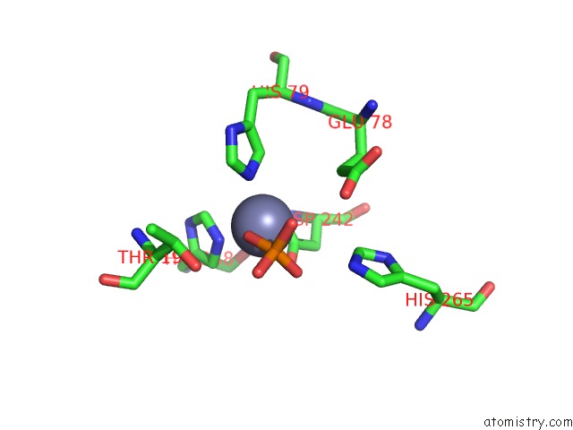
Zinc binding site 4 out of 4 in 4mdt
Go back to
Zinc binding site 4 out
of 4 in the Structure of Lpxc Bound to the Reaction Product Udp-(3-O-(R-3- Hydroxymyristoyl))-Glucosamine

Mono view

Stereo pair view
Mono view
Stereo pair view
A full contact list of Zinc with other atoms in the Zn binding
site number 4 of Structure of Lpxc Bound to the Reaction Product Udp-(3-O-(R-3- Hydroxymyristoyl))-Glucosamine within 5.0Å range:
|
Reference:
G.M.Clayton,
D.J.Klein,
K.W.Rickert,
S.B.Patel,
M.Kornienko,
J.Zugay-Murphy,
J.C.Reid,
S.Tummala,
S.Sharma,
S.B.Singh,
L.Miesel,
K.J.Lumb,
S.M.Soisson.
Structure of the Bacterial Deacetylase Lpxc Bound to the Nucleotide Reaction Product Reveals Mechanisms of Oxyanion Stabilization and Proton Transfer. J.Biol.Chem. V. 288 34073 2013.
ISSN: ISSN 0021-9258
PubMed: 24108127
DOI: 10.1074/JBC.M113.513028
Page generated: Sun Oct 27 02:23:23 2024
ISSN: ISSN 0021-9258
PubMed: 24108127
DOI: 10.1074/JBC.M113.513028
Last articles
Zn in 9J0NZn in 9J0O
Zn in 9J0P
Zn in 9FJX
Zn in 9EKB
Zn in 9C0F
Zn in 9CAH
Zn in 9CH0
Zn in 9CH3
Zn in 9CH1