Zinc in PDB 4kis: Crystal Structure of A Lsr-Dna Complex
Protein crystallography data
The structure of Crystal Structure of A Lsr-Dna Complex, PDB code: 4kis
was solved by
K.Rutherford,
P.Yuan,
K.Perry,
G.D.Van Duyne,
with X-Ray Crystallography technique. A brief refinement statistics is given in the table below:
Resolution Low / High (Å)
|
49.86 /
3.20
|
Space group
|
I 2 3
|
Cell size a, b, c (Å), α, β, γ (°)
|
290.750,
290.750,
290.750,
90.00,
90.00,
90.00
|
R / Rfree (%)
|
23.6 /
25.6
|
Other elements in 4kis:
The structure of Crystal Structure of A Lsr-Dna Complex also contains other interesting chemical elements:
Zinc Binding Sites:
The binding sites of Zinc atom in the Crystal Structure of A Lsr-Dna Complex
(pdb code 4kis). This binding sites where shown within
5.0 Angstroms radius around Zinc atom.
In total 6 binding sites of Zinc where determined in the
Crystal Structure of A Lsr-Dna Complex, PDB code: 4kis:
Jump to Zinc binding site number:
1;
2;
3;
4;
5;
6;
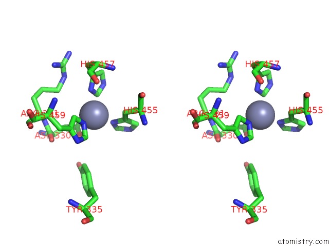
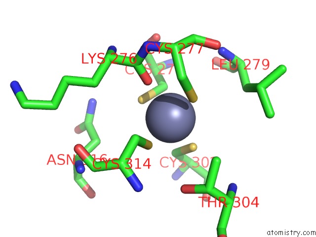
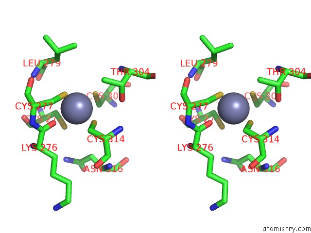
Zinc binding site 1 out
of 6 in 4kis
Go back to
Zinc Binding Sites List in 4kis
Zinc binding site 1 out
of 6 in the Crystal Structure of A Lsr-Dna Complex
 Mono view
 Stereo pair view
|
A full contact list of Zinc with other atoms in the Zn binding
site number 1 of Crystal Structure of A Lsr-Dna Complex within 5.0Å range:
probe
|
atom
|
residue
|
distance (Å)
|
B
|
Occ
|
A:Zn501
b:0.8
occ:1.00
|
SG
|
A:CYS302
|
2.3
|
81.9
|
1.0
|
SG
|
A:CYS274
|
2.3
|
96.9
|
1.0
|
SG
|
A:CYS277
|
2.3
|
96.6
|
1.0
|
O
|
A:LYS276
|
2.3
|
97.1
|
1.0
|
SG
|
A:CYS314
|
2.4
|
0.0
|
1.0
|
CB
|
A:CYS274
|
2.9
|
96.9
|
1.0
|
CB
|
A:CYS302
|
3.4
|
81.9
|
1.0
|
C
|
A:LYS276
|
3.4
|
97.1
|
1.0
|
CB
|
A:CYS314
|
3.4
|
0.0
|
1.0
|
CB
|
A:CYS277
|
3.6
|
96.6
|
1.0
|
OG1
|
A:THR304
|
4.0
|
0.1
|
1.0
|
CB
|
A:LYS276
|
4.2
|
0.8
|
1.0
|
N
|
A:CYS277
|
4.3
|
0.4
|
1.0
|
CA
|
A:LYS276
|
4.3
|
97.1
|
1.0
|
CA
|
A:CYS274
|
4.4
|
93.9
|
1.0
|
CA
|
A:CYS277
|
4.4
|
0.4
|
1.0
|
CG2
|
A:THR304
|
4.4
|
0.1
|
1.0
|
N
|
A:LYS276
|
4.6
|
97.1
|
1.0
|
N
|
A:GLY278
|
4.7
|
92.7
|
1.0
|
CA
|
A:CYS302
|
4.7
|
78.3
|
1.0
|
C
|
A:CYS277
|
4.7
|
0.4
|
1.0
|
C
|
A:CYS274
|
4.8
|
93.9
|
1.0
|
O
|
A:CYS274
|
4.8
|
93.9
|
1.0
|
CB
|
A:THR304
|
4.8
|
0.1
|
1.0
|
CB
|
A:ASN316
|
4.9
|
0.1
|
1.0
|
N
|
A:LEU279
|
4.9
|
90.9
|
1.0
|
CA
|
A:CYS314
|
4.9
|
0.8
|
1.0
|
O
|
A:LEU279
|
4.9
|
90.9
|
1.0
|
|
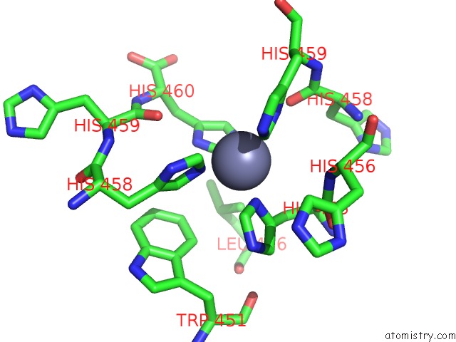
Zinc binding site 2 out
of 6 in 4kis
Go back to
Zinc Binding Sites List in 4kis
Zinc binding site 2 out
of 6 in the Crystal Structure of A Lsr-Dna Complex
 Mono view
 Stereo pair view
|
A full contact list of Zinc with other atoms in the Zn binding
site number 2 of Crystal Structure of A Lsr-Dna Complex within 5.0Å range:
probe
|
atom
|
residue
|
distance (Å)
|
B
|
Occ
|
A:Zn502
b:87.5
occ:1.00
|
ND1
|
A:HIS455
|
2.0
|
94.7
|
1.0
|
OD2
|
D:ASP330
|
2.1
|
81.3
|
1.0
|
CE1
|
A:HIS457
|
2.1
|
0.1
|
1.0
|
NE2
|
A:HIS459
|
2.2
|
97.1
|
1.0
|
ND1
|
A:HIS457
|
2.4
|
0.1
|
1.0
|
CG
|
D:ASP330
|
2.9
|
81.3
|
1.0
|
CG
|
A:HIS455
|
3.0
|
94.7
|
1.0
|
OD1
|
D:ASP330
|
3.0
|
81.3
|
1.0
|
CE1
|
A:HIS459
|
3.1
|
97.1
|
1.0
|
CE1
|
A:HIS455
|
3.1
|
94.7
|
1.0
|
CB
|
A:HIS455
|
3.2
|
94.7
|
1.0
|
NE2
|
A:HIS457
|
3.4
|
0.1
|
1.0
|
CD2
|
A:HIS459
|
3.4
|
97.1
|
1.0
|
CG
|
A:HIS457
|
3.8
|
0.1
|
1.0
|
CD2
|
A:HIS455
|
4.2
|
94.7
|
1.0
|
CD2
|
A:HIS457
|
4.2
|
0.1
|
1.0
|
CD
|
D:ARG331
|
4.2
|
86.5
|
1.0
|
NE2
|
A:HIS455
|
4.2
|
94.7
|
1.0
|
CB
|
D:ASP330
|
4.3
|
81.3
|
1.0
|
ND1
|
A:HIS459
|
4.3
|
97.1
|
1.0
|
CG
|
A:HIS459
|
4.5
|
97.1
|
1.0
|
O
|
D:ASP330
|
4.6
|
82.6
|
1.0
|
C
|
D:ASP330
|
4.7
|
82.6
|
1.0
|
NE
|
D:ARG331
|
4.7
|
86.5
|
1.0
|
CA
|
A:HIS455
|
4.7
|
89.1
|
1.0
|
N
|
D:ARG331
|
4.9
|
89.2
|
1.0
|
CB
|
A:HIS457
|
4.9
|
0.1
|
1.0
|
CE2
|
D:TYR335
|
4.9
|
0.3
|
1.0
|
|
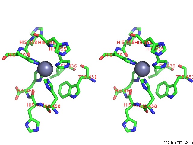
Zinc binding site 3 out
of 6 in 4kis
Go back to
Zinc Binding Sites List in 4kis
Zinc binding site 3 out
of 6 in the Crystal Structure of A Lsr-Dna Complex
 Mono view
 Stereo pair view
|
A full contact list of Zinc with other atoms in the Zn binding
site number 3 of Crystal Structure of A Lsr-Dna Complex within 5.0Å range:
probe
|
atom
|
residue
|
distance (Å)
|
B
|
Occ
|
A:Zn503
b:0.8
occ:1.00
|
CE1
|
A:HIS460
|
2.0
|
86.7
|
1.0
|
NE2
|
A:HIS458
|
2.1
|
0.9
|
1.0
|
ND1
|
D:HIS455
|
2.2
|
0.2
|
1.0
|
CE1
|
D:HIS459
|
2.5
|
0.7
|
1.0
|
NE2
|
A:HIS460
|
2.8
|
86.7
|
1.0
|
CE1
|
A:HIS458
|
2.9
|
0.9
|
1.0
|
ND1
|
D:HIS459
|
3.0
|
0.7
|
1.0
|
ND1
|
A:HIS460
|
3.1
|
86.7
|
1.0
|
CE1
|
D:HIS455
|
3.1
|
0.2
|
1.0
|
CG
|
D:HIS455
|
3.2
|
0.2
|
1.0
|
CD2
|
A:HIS458
|
3.3
|
0.9
|
1.0
|
CB
|
D:HIS455
|
3.6
|
0.2
|
1.0
|
NE2
|
D:HIS459
|
3.6
|
0.7
|
1.0
|
O
|
D:HIS458
|
4.0
|
0.5
|
1.0
|
CD2
|
A:HIS460
|
4.0
|
86.7
|
1.0
|
CG
|
A:HIS460
|
4.1
|
86.7
|
1.0
|
ND1
|
A:HIS458
|
4.2
|
0.9
|
1.0
|
N
|
D:HIS456
|
4.2
|
0.0
|
1.0
|
NE2
|
D:HIS455
|
4.3
|
0.2
|
1.0
|
CG
|
D:HIS459
|
4.3
|
0.7
|
1.0
|
CA
|
D:HIS455
|
4.3
|
0.7
|
1.0
|
CD2
|
D:HIS455
|
4.3
|
0.2
|
1.0
|
CG
|
A:HIS458
|
4.3
|
0.9
|
1.0
|
C
|
D:HIS455
|
4.4
|
0.7
|
1.0
|
O
|
A:HIS459
|
4.5
|
87.5
|
1.0
|
CD2
|
D:HIS459
|
4.6
|
0.7
|
1.0
|
CD1
|
D:LEU436
|
4.7
|
82.7
|
1.0
|
CZ3
|
D:TRP451
|
4.9
|
81.8
|
1.0
|
CA
|
D:HIS456
|
4.9
|
0.0
|
1.0
|
CE3
|
D:TRP451
|
4.9
|
81.8
|
1.0
|
C
|
D:HIS458
|
5.0
|
0.5
|
1.0
|
|
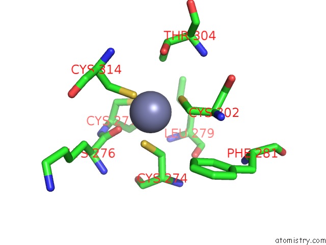
Zinc binding site 4 out
of 6 in 4kis
Go back to
Zinc Binding Sites List in 4kis
Zinc binding site 4 out
of 6 in the Crystal Structure of A Lsr-Dna Complex
 Mono view
 Stereo pair view
|
A full contact list of Zinc with other atoms in the Zn binding
site number 4 of Crystal Structure of A Lsr-Dna Complex within 5.0Å range:
probe
|
atom
|
residue
|
distance (Å)
|
B
|
Occ
|
B:Zn501
b:94.5
occ:1.00
|
SG
|
B:CYS302
|
2.2
|
70.5
|
1.0
|
SG
|
B:CYS274
|
2.3
|
89.1
|
1.0
|
SG
|
B:CYS277
|
2.3
|
80.6
|
1.0
|
SG
|
B:CYS314
|
2.3
|
96.5
|
1.0
|
O
|
B:LYS276
|
2.6
|
82.8
|
1.0
|
CB
|
B:CYS274
|
3.0
|
89.1
|
1.0
|
CB
|
B:CYS302
|
3.2
|
70.5
|
1.0
|
CB
|
B:CYS314
|
3.4
|
96.5
|
1.0
|
CG2
|
B:THR304
|
3.7
|
81.5
|
1.0
|
CB
|
B:CYS277
|
3.7
|
80.6
|
1.0
|
C
|
B:LYS276
|
3.9
|
82.8
|
1.0
|
OG1
|
B:THR304
|
4.3
|
81.5
|
1.0
|
CA
|
B:CYS274
|
4.5
|
76.2
|
1.0
|
CA
|
B:CYS302
|
4.6
|
68.1
|
1.0
|
CA
|
B:CYS277
|
4.6
|
82.0
|
1.0
|
C
|
B:CYS277
|
4.7
|
82.0
|
1.0
|
N
|
B:CYS277
|
4.7
|
82.0
|
1.0
|
O
|
B:CYS277
|
4.7
|
82.0
|
1.0
|
CB
|
B:THR304
|
4.7
|
81.5
|
1.0
|
CA
|
B:CYS314
|
4.8
|
0.9
|
1.0
|
N
|
B:LEU279
|
4.8
|
78.0
|
1.0
|
CB
|
B:LEU279
|
4.8
|
79.2
|
1.0
|
CA
|
B:LYS276
|
4.9
|
82.8
|
1.0
|
CB
|
B:LYS276
|
4.9
|
88.6
|
1.0
|
O
|
B:LEU279
|
4.9
|
78.0
|
1.0
|
CE2
|
B:PHE281
|
5.0
|
84.3
|
1.0
|
|
Zinc binding site 5 out
of 6 in 4kis
Go back to
Zinc Binding Sites List in 4kis
Zinc binding site 5 out
of 6 in the Crystal Structure of A Lsr-Dna Complex
 Mono view
 Stereo pair view
|
A full contact list of Zinc with other atoms in the Zn binding
site number 5 of Crystal Structure of A Lsr-Dna Complex within 5.0Å range:
probe
|
atom
|
residue
|
distance (Å)
|
B
|
Occ
|
C:Zn501
b:0.1
occ:1.00
|
SG
|
C:CYS277
|
2.3
|
0.8
|
1.0
|
SG
|
C:CYS314
|
2.3
|
0.8
|
1.0
|
SG
|
C:CYS302
|
2.4
|
0.7
|
1.0
|
SG
|
C:CYS274
|
2.4
|
0.3
|
1.0
|
O
|
C:LYS276
|
2.6
|
0.2
|
1.0
|
CB
|
C:CYS274
|
2.8
|
0.3
|
1.0
|
CB
|
C:CYS302
|
3.5
|
0.7
|
1.0
|
CB
|
C:CYS314
|
3.6
|
0.8
|
1.0
|
CB
|
C:CYS277
|
3.6
|
0.8
|
1.0
|
C
|
C:LYS276
|
3.7
|
0.2
|
1.0
|
CG2
|
C:THR304
|
4.0
|
0.5
|
1.0
|
CA
|
C:CYS274
|
4.3
|
0.5
|
1.0
|
CA
|
C:CYS277
|
4.5
|
0.7
|
1.0
|
N
|
C:CYS277
|
4.5
|
0.7
|
1.0
|
O
|
C:CYS274
|
4.5
|
0.5
|
1.0
|
OG1
|
C:THR304
|
4.6
|
0.5
|
1.0
|
CB
|
C:LYS276
|
4.6
|
0.9
|
1.0
|
N
|
C:GLY278
|
4.6
|
1.0
|
1.0
|
CA
|
C:LYS276
|
4.7
|
0.2
|
1.0
|
C
|
C:CYS274
|
4.7
|
0.5
|
1.0
|
O
|
C:LEU279
|
4.8
|
0.9
|
1.0
|
N
|
C:LEU279
|
4.8
|
0.9
|
1.0
|
C
|
C:CYS277
|
4.8
|
0.7
|
1.0
|
CA
|
C:CYS302
|
4.9
|
0.5
|
1.0
|
N
|
C:LYS276
|
4.9
|
0.2
|
1.0
|
CB
|
C:THR304
|
4.9
|
0.5
|
1.0
|
CA
|
C:CYS314
|
4.9
|
0.3
|
1.0
|
|
Zinc binding site 6 out
of 6 in 4kis
Go back to
Zinc Binding Sites List in 4kis
Zinc binding site 6 out
of 6 in the Crystal Structure of A Lsr-Dna Complex
 Mono view
 Stereo pair view
|
A full contact list of Zinc with other atoms in the Zn binding
site number 6 of Crystal Structure of A Lsr-Dna Complex within 5.0Å range:
probe
|
atom
|
residue
|
distance (Å)
|
B
|
Occ
|
D:Zn501
b:0.4
occ:1.00
|
SG
|
D:CYS274
|
2.3
|
94.0
|
1.0
|
SG
|
D:CYS314
|
2.3
|
0.8
|
1.0
|
SG
|
D:CYS302
|
2.3
|
84.1
|
1.0
|
SG
|
D:CYS277
|
2.4
|
97.6
|
1.0
|
O
|
D:LYS276
|
2.6
|
0.1
|
1.0
|
CB
|
D:CYS274
|
3.0
|
94.0
|
1.0
|
CB
|
D:CYS302
|
3.2
|
84.1
|
1.0
|
CB
|
D:CYS314
|
3.6
|
0.8
|
1.0
|
C
|
D:LYS276
|
3.8
|
0.1
|
1.0
|
CG2
|
D:THR304
|
3.9
|
0.9
|
1.0
|
CB
|
D:CYS277
|
3.9
|
97.6
|
1.0
|
OG1
|
D:THR304
|
4.1
|
0.9
|
1.0
|
CA
|
D:CYS274
|
4.4
|
91.1
|
1.0
|
CA
|
D:CYS302
|
4.6
|
90.0
|
1.0
|
CB
|
D:LEU279
|
4.6
|
85.0
|
1.0
|
CA
|
D:CYS277
|
4.7
|
0.6
|
1.0
|
CB
|
D:THR304
|
4.7
|
0.9
|
1.0
|
N
|
D:CYS277
|
4.7
|
0.6
|
1.0
|
CB
|
D:LYS276
|
4.7
|
0.3
|
1.0
|
N
|
D:LEU279
|
4.7
|
85.2
|
1.0
|
C
|
D:CYS277
|
4.7
|
0.6
|
1.0
|
CA
|
D:LYS276
|
4.8
|
0.1
|
1.0
|
CB
|
D:ASN316
|
4.8
|
0.1
|
1.0
|
C
|
D:CYS274
|
4.9
|
91.1
|
1.0
|
O
|
D:CYS274
|
4.9
|
91.1
|
1.0
|
O
|
D:CYS277
|
4.9
|
0.6
|
1.0
|
N
|
D:LYS276
|
5.0
|
0.1
|
1.0
|
CA
|
D:CYS314
|
5.0
|
0.6
|
1.0
|
|
Reference:
K.Rutherford,
P.Yuan,
K.Perry,
R.Sharp,
G.D.Van Duyne.
Attachment Site Recognition and Regulation of Directionality By the Serine Integrases. Nucleic Acids Res. V. 41 8341 2013.
ISSN: ISSN 0305-1048
PubMed: 23821671
DOI: 10.1093/NAR/GKT580
Page generated: Sun Oct 27 01:14:09 2024
|