Zinc »
PDB 4i9i-4ii1 »
4if6 »
Zinc in PDB 4if6: Structure of Nad-Dependent Protein Deacetylase Sirtuin-1 (Closed State, 2.25 A)
Protein crystallography data
The structure of Structure of Nad-Dependent Protein Deacetylase Sirtuin-1 (Closed State, 2.25 A), PDB code: 4if6
was solved by
A.M.Davenport,
F.M.Huber,
A.Hoelz,
with X-Ray Crystallography technique. A brief refinement statistics is given in the table below:
| Resolution Low / High (Å) | 19.65 / 2.25 |
| Space group | P 32 2 1 |
| Cell size a, b, c (Å), α, β, γ (°) | 92.662, 92.662, 97.700, 90.00, 90.00, 120.00 |
| R / Rfree (%) | 15.6 / 18.7 |
Zinc Binding Sites:
The binding sites of Zinc atom in the Structure of Nad-Dependent Protein Deacetylase Sirtuin-1 (Closed State, 2.25 A)
(pdb code 4if6). This binding sites where shown within
5.0 Angstroms radius around Zinc atom.
In total only one binding site of Zinc was determined in the Structure of Nad-Dependent Protein Deacetylase Sirtuin-1 (Closed State, 2.25 A), PDB code: 4if6:
In total only one binding site of Zinc was determined in the Structure of Nad-Dependent Protein Deacetylase Sirtuin-1 (Closed State, 2.25 A), PDB code: 4if6:
Zinc binding site 1 out of 1 in 4if6
Go back to
Zinc binding site 1 out
of 1 in the Structure of Nad-Dependent Protein Deacetylase Sirtuin-1 (Closed State, 2.25 A)
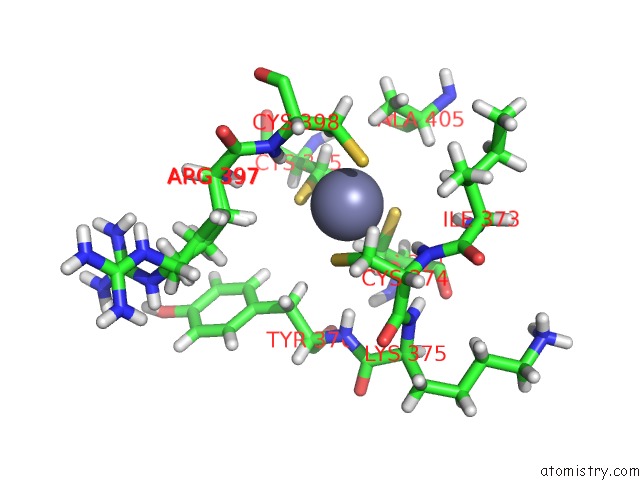
Mono view
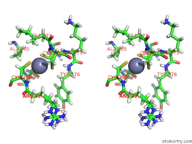
Stereo pair view
Mono view
Stereo pair view
A full contact list of Zinc with other atoms in the Zn binding
site number 1 of Structure of Nad-Dependent Protein Deacetylase Sirtuin-1 (Closed State, 2.25 A) within 5.0Å range:
|
Reference:
A.M.Davenport,
F.M.Huber,
A.Hoelz.
Structure of A Nucleoporin Complex To Be Published.
Page generated: Sun Oct 27 00:42:44 2024
Last articles
Zn in 9J0NZn in 9J0O
Zn in 9J0P
Zn in 9FJX
Zn in 9EKB
Zn in 9C0F
Zn in 9CAH
Zn in 9CH0
Zn in 9CH3
Zn in 9CH1