Zinc »
PDB 4hwt-4i7h »
4i1h »
Zinc in PDB 4i1h: Structure of Parkin E3 Ligase
Protein crystallography data
The structure of Structure of Parkin E3 Ligase, PDB code: 4i1h
was solved by
J.C.Lougheed,
E.Brecht,
N.H.Yao,
with X-Ray Crystallography technique. A brief refinement statistics is given in the table below:
| Resolution Low / High (Å) | 16.97 / 2.00 |
| Space group | C 2 2 21 |
| Cell size a, b, c (Å), α, β, γ (°) | 87.110, 133.930, 66.210, 90.00, 90.00, 90.00 |
| R / Rfree (%) | 18.1 / 21.5 |
Zinc Binding Sites:
The binding sites of Zinc atom in the Structure of Parkin E3 Ligase
(pdb code 4i1h). This binding sites where shown within
5.0 Angstroms radius around Zinc atom.
In total 8 binding sites of Zinc where determined in the Structure of Parkin E3 Ligase, PDB code: 4i1h:
Jump to Zinc binding site number: 1; 2; 3; 4; 5; 6; 7; 8;
In total 8 binding sites of Zinc where determined in the Structure of Parkin E3 Ligase, PDB code: 4i1h:
Jump to Zinc binding site number: 1; 2; 3; 4; 5; 6; 7; 8;
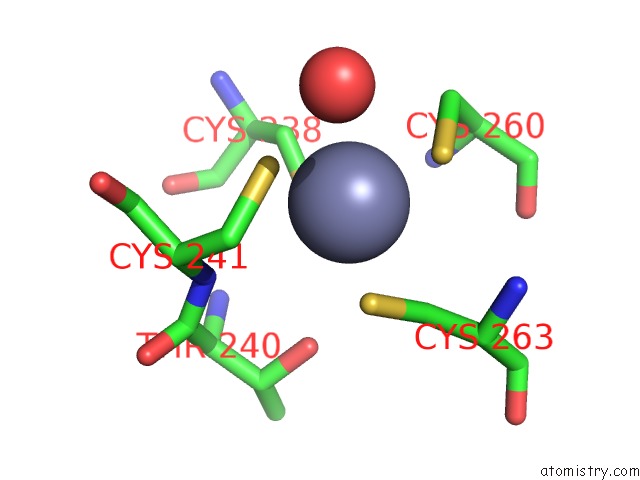
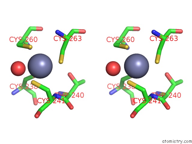
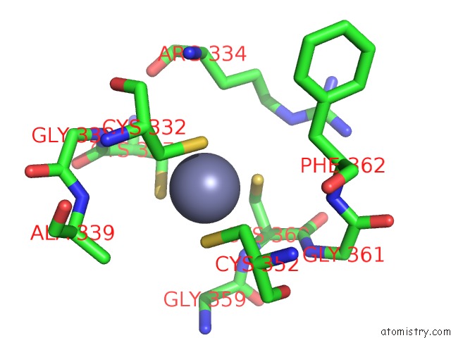
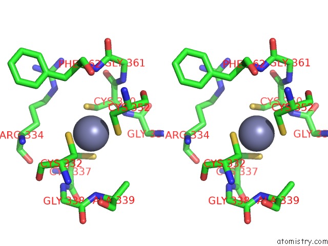
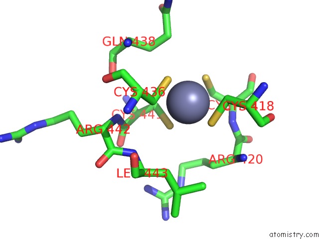
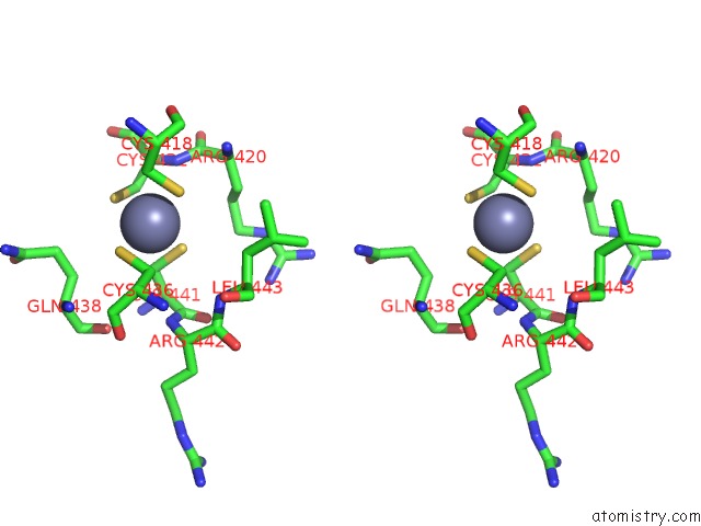
Zinc binding site 1 out of 8 in 4i1h
Go back to
Zinc binding site 1 out
of 8 in the Structure of Parkin E3 Ligase
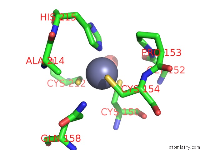
Mono view
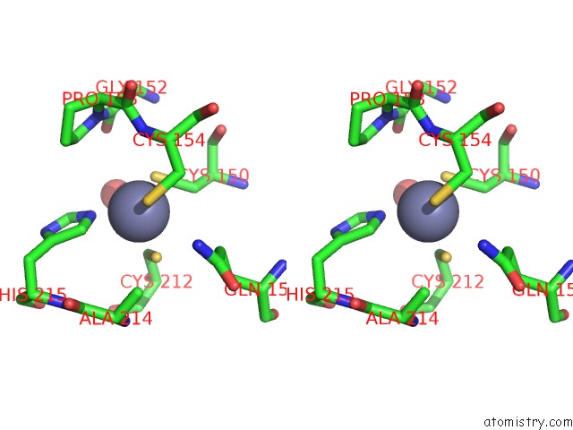
Stereo pair view
Mono view
Stereo pair view
A full contact list of Zinc with other atoms in the Zn binding
site number 1 of Structure of Parkin E3 Ligase within 5.0Å range:
|
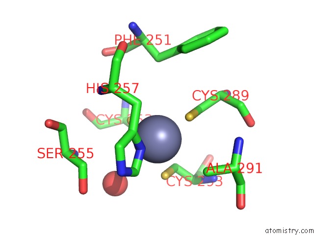
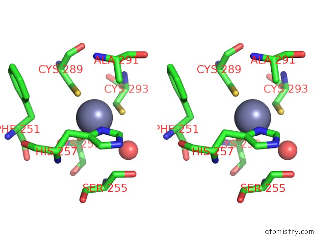
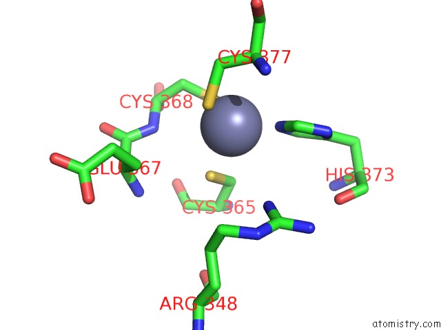
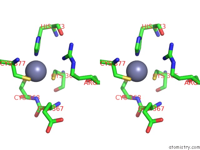
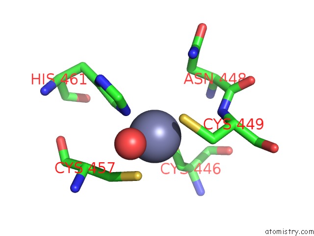
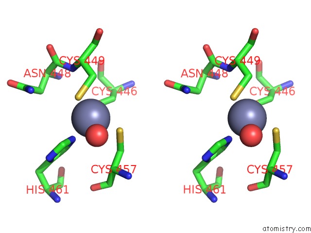
Zinc binding site 2 out of 8 in 4i1h
Go back to
Zinc binding site 2 out
of 8 in the Structure of Parkin E3 Ligase
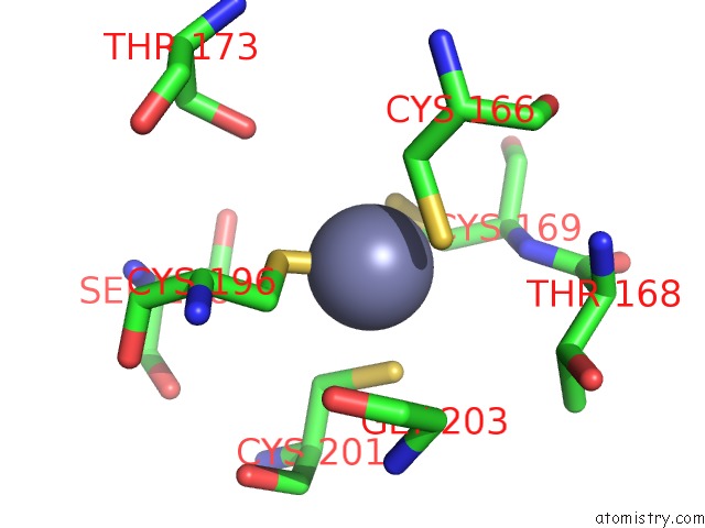
Mono view
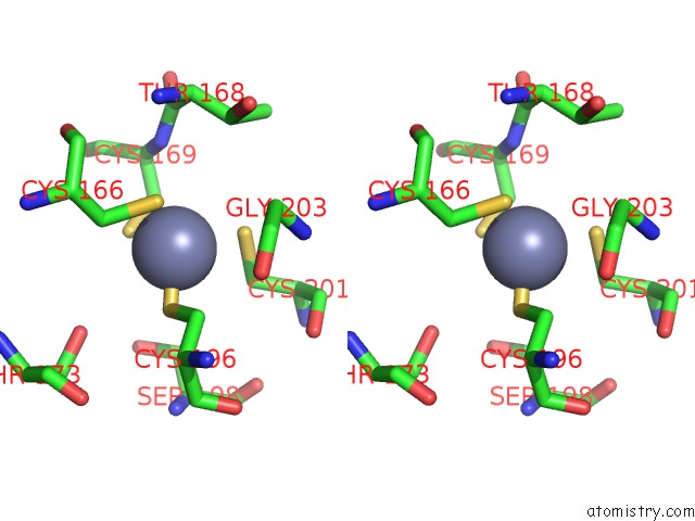
Stereo pair view
Mono view
Stereo pair view
A full contact list of Zinc with other atoms in the Zn binding
site number 2 of Structure of Parkin E3 Ligase within 5.0Å range:
|
Zinc binding site 3 out of 8 in 4i1h
Go back to
Zinc binding site 3 out
of 8 in the Structure of Parkin E3 Ligase
Mono view
Stereo pair view
Mono view
Stereo pair view
A full contact list of Zinc with other atoms in the Zn binding
site number 3 of Structure of Parkin E3 Ligase within 5.0Å range:
|
Zinc binding site 4 out of 8 in 4i1h
Go back to
Zinc binding site 4 out
of 8 in the Structure of Parkin E3 Ligase
Mono view
Stereo pair view
Mono view
Stereo pair view
A full contact list of Zinc with other atoms in the Zn binding
site number 4 of Structure of Parkin E3 Ligase within 5.0Å range:
|
Zinc binding site 5 out of 8 in 4i1h
Go back to
Zinc binding site 5 out
of 8 in the Structure of Parkin E3 Ligase
Mono view
Stereo pair view
Mono view
Stereo pair view
A full contact list of Zinc with other atoms in the Zn binding
site number 5 of Structure of Parkin E3 Ligase within 5.0Å range:
|
Zinc binding site 6 out of 8 in 4i1h
Go back to
Zinc binding site 6 out
of 8 in the Structure of Parkin E3 Ligase
Mono view
Stereo pair view
Mono view
Stereo pair view
A full contact list of Zinc with other atoms in the Zn binding
site number 6 of Structure of Parkin E3 Ligase within 5.0Å range:
|
Zinc binding site 7 out of 8 in 4i1h
Go back to
Zinc binding site 7 out
of 8 in the Structure of Parkin E3 Ligase
Mono view
Stereo pair view
Mono view
Stereo pair view
A full contact list of Zinc with other atoms in the Zn binding
site number 7 of Structure of Parkin E3 Ligase within 5.0Å range:
|
Zinc binding site 8 out of 8 in 4i1h
Go back to
Zinc binding site 8 out
of 8 in the Structure of Parkin E3 Ligase
Mono view
Stereo pair view
Mono view
Stereo pair view
A full contact list of Zinc with other atoms in the Zn binding
site number 8 of Structure of Parkin E3 Ligase within 5.0Å range:
|
Reference:
B.E.Riley,
J.C.Lougheed,
K.Callaway,
M.Velasquez,
E.Brecht,
L.Nguyen,
T.Shaler,
D.Walker,
Y.Yang,
K.Regnstrom,
L.Diep,
Z.Zhang,
S.Chiou,
M.Bova,
D.R.Artis,
N.Yao,
J.Baker,
T.Yednock,
J.A.Johnston.
Structure and Function of Parkin E3 Ubiquitin Ligase Reveals Aspects of Ring and Hect Ligases. Nat Commun V. 4 1982 2013.
ISSN: ESSN 2041-1723
PubMed: 23770887
DOI: 10.1038/NCOMMS2982
Page generated: Sun Oct 27 00:27:13 2024
ISSN: ESSN 2041-1723
PubMed: 23770887
DOI: 10.1038/NCOMMS2982
Last articles
Zn in 9J0NZn in 9J0O
Zn in 9J0P
Zn in 9FJX
Zn in 9EKB
Zn in 9C0F
Zn in 9CAH
Zn in 9CH0
Zn in 9CH3
Zn in 9CH1