Zinc »
PDB 4fvw-4g2z »
4fyt »
Zinc in PDB 4fyt: Human Aminopeptidase N (CD13) in Complex with Amastatin
Enzymatic activity of Human Aminopeptidase N (CD13) in Complex with Amastatin
All present enzymatic activity of Human Aminopeptidase N (CD13) in Complex with Amastatin:
3.4.11.2;
3.4.11.2;
Protein crystallography data
The structure of Human Aminopeptidase N (CD13) in Complex with Amastatin, PDB code: 4fyt
was solved by
A.H.Wong,
J.M.Rini,
with X-Ray Crystallography technique. A brief refinement statistics is given in the table below:
| Resolution Low / High (Å) | 47.04 / 1.85 |
| Space group | P 64 |
| Cell size a, b, c (Å), α, β, γ (°) | 157.470, 157.470, 115.130, 90.00, 90.00, 120.00 |
| R / Rfree (%) | 16.9 / 18.3 |
Zinc Binding Sites:
The binding sites of Zinc atom in the Human Aminopeptidase N (CD13) in Complex with Amastatin
(pdb code 4fyt). This binding sites where shown within
5.0 Angstroms radius around Zinc atom.
In total only one binding site of Zinc was determined in the Human Aminopeptidase N (CD13) in Complex with Amastatin, PDB code: 4fyt:
In total only one binding site of Zinc was determined in the Human Aminopeptidase N (CD13) in Complex with Amastatin, PDB code: 4fyt:
Zinc binding site 1 out of 1 in 4fyt
Go back to
Zinc binding site 1 out
of 1 in the Human Aminopeptidase N (CD13) in Complex with Amastatin
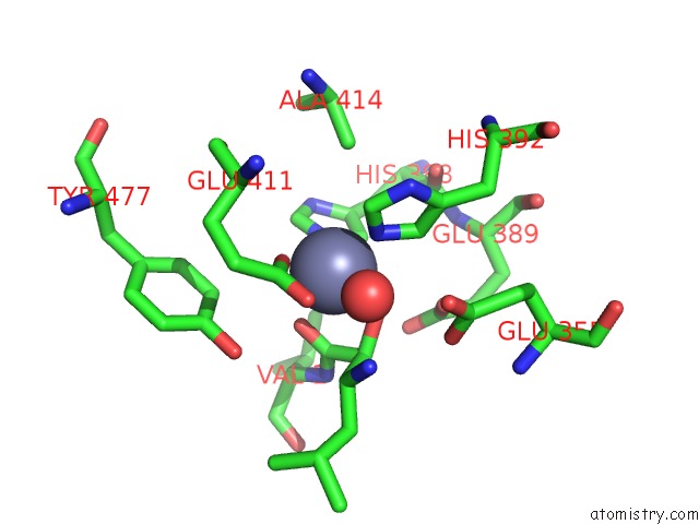
Mono view
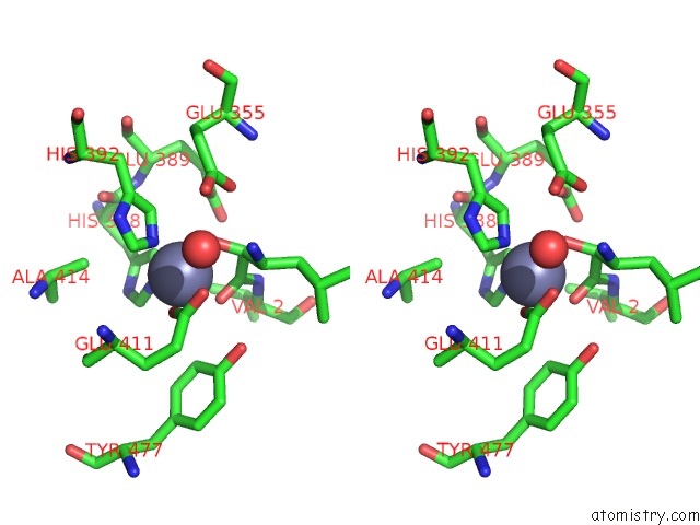
Stereo pair view
Mono view
Stereo pair view
A full contact list of Zinc with other atoms in the Zn binding
site number 1 of Human Aminopeptidase N (CD13) in Complex with Amastatin within 5.0Å range:
|
Reference:
A.H.Wong,
D.Zhou,
J.M.Rini.
The X-Ray Crystal Structure of Human Aminopeptidase N Reveals A Novel Dimer and the Basis For Peptide Processing. J.Biol.Chem. V. 287 36804 2012.
ISSN: ISSN 0021-9258
PubMed: 22932899
DOI: 10.1074/JBC.M112.398842
Page generated: Sat Oct 26 23:02:05 2024
ISSN: ISSN 0021-9258
PubMed: 22932899
DOI: 10.1074/JBC.M112.398842
Last articles
Zn in 9J0NZn in 9J0O
Zn in 9J0P
Zn in 9FJX
Zn in 9EKB
Zn in 9C0F
Zn in 9CAH
Zn in 9CH0
Zn in 9CH3
Zn in 9CH1