Zinc »
PDB 4eyp-4f6z »
4f2j »
Zinc in PDB 4f2j: Crystal Structure of ZNF217 Bound to Dna, P6522 Crystal Form
Protein crystallography data
The structure of Crystal Structure of ZNF217 Bound to Dna, P6522 Crystal Form, PDB code: 4f2j
was solved by
M.S.Vandevenne,
D.A.Jacques,
J.M.Guss,
J.P.Mackay,
with X-Ray Crystallography technique. A brief refinement statistics is given in the table below:
| Resolution Low / High (Å) | 48.31 / 2.64 |
| Space group | P 65 2 2 |
| Cell size a, b, c (Å), α, β, γ (°) | 56.928, 56.928, 242.542, 90.00, 90.00, 120.00 |
| R / Rfree (%) | 25.2 / 26.4 |
Zinc Binding Sites:
The binding sites of Zinc atom in the Crystal Structure of ZNF217 Bound to Dna, P6522 Crystal Form
(pdb code 4f2j). This binding sites where shown within
5.0 Angstroms radius around Zinc atom.
In total 2 binding sites of Zinc where determined in the Crystal Structure of ZNF217 Bound to Dna, P6522 Crystal Form, PDB code: 4f2j:
Jump to Zinc binding site number: 1; 2;
In total 2 binding sites of Zinc where determined in the Crystal Structure of ZNF217 Bound to Dna, P6522 Crystal Form, PDB code: 4f2j:
Jump to Zinc binding site number: 1; 2;
Zinc binding site 1 out of 2 in 4f2j
Go back to
Zinc binding site 1 out
of 2 in the Crystal Structure of ZNF217 Bound to Dna, P6522 Crystal Form

Mono view
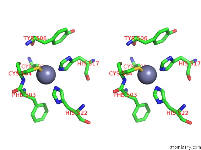
Stereo pair view
Mono view
Stereo pair view
A full contact list of Zinc with other atoms in the Zn binding
site number 1 of Crystal Structure of ZNF217 Bound to Dna, P6522 Crystal Form within 5.0Å range:
|
Zinc binding site 2 out of 2 in 4f2j
Go back to
Zinc binding site 2 out
of 2 in the Crystal Structure of ZNF217 Bound to Dna, P6522 Crystal Form

Mono view

Stereo pair view
Mono view
Stereo pair view
A full contact list of Zinc with other atoms in the Zn binding
site number 2 of Crystal Structure of ZNF217 Bound to Dna, P6522 Crystal Form within 5.0Å range:
|
Reference:
M.S.Vandevenne,
D.A.Jacques,
C.Artuz,
C.D.Nguyen,
A.H.Kwan,
D.J.Segal,
J.M.Matthews,
M.Crossley,
J.M.Guss,
J.P.Mackay.
New Insights Into Dna Recognition By Zinc Fingers Revealed By Structural Analysis of the Oncoprotein ZNF217 J.Biol.Chem. V. 288 10616 2013.
ISSN: ISSN 0021-9258
PubMed: 23436653
DOI: 10.1074/JBC.M112.441451
Page generated: Sat Oct 26 22:15:27 2024
ISSN: ISSN 0021-9258
PubMed: 23436653
DOI: 10.1074/JBC.M112.441451
Last articles
Zn in 9J0NZn in 9J0O
Zn in 9J0P
Zn in 9FJX
Zn in 9EKB
Zn in 9C0F
Zn in 9CAH
Zn in 9CH0
Zn in 9CH3
Zn in 9CH1