Zinc »
PDB 4eyp-4f6z »
4f29 »
Zinc in PDB 4f29: Quisqualate Bound to the Ligand Binding Domain of GLUA3I
Protein crystallography data
The structure of Quisqualate Bound to the Ligand Binding Domain of GLUA3I, PDB code: 4f29
was solved by
A.H.Ahmed,
R.E.Oswald,
with X-Ray Crystallography technique. A brief refinement statistics is given in the table below:
| Resolution Low / High (Å) | 44.83 / 1.75 |
| Space group | P 2 2 21 |
| Cell size a, b, c (Å), α, β, γ (°) | 47.411, 47.516, 137.595, 90.00, 90.00, 90.00 |
| R / Rfree (%) | 18.8 / 24 |
Zinc Binding Sites:
The binding sites of Zinc atom in the Quisqualate Bound to the Ligand Binding Domain of GLUA3I
(pdb code 4f29). This binding sites where shown within
5.0 Angstroms radius around Zinc atom.
In total only one binding site of Zinc was determined in the Quisqualate Bound to the Ligand Binding Domain of GLUA3I, PDB code: 4f29:
In total only one binding site of Zinc was determined in the Quisqualate Bound to the Ligand Binding Domain of GLUA3I, PDB code: 4f29:
Zinc binding site 1 out of 1 in 4f29
Go back to
Zinc binding site 1 out
of 1 in the Quisqualate Bound to the Ligand Binding Domain of GLUA3I
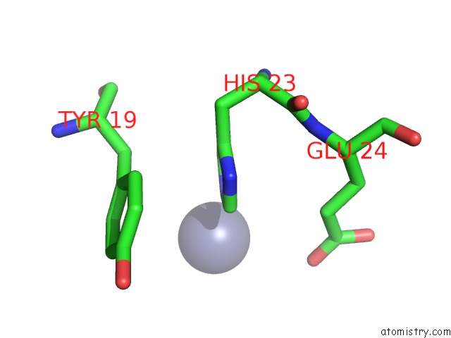
Mono view
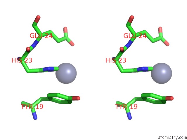
Stereo pair view
Mono view
Stereo pair view
A full contact list of Zinc with other atoms in the Zn binding
site number 1 of Quisqualate Bound to the Ligand Binding Domain of GLUA3I within 5.0Å range:
|
Reference:
S.M.Holley,
A.H.Ahmed,
J.Srinivasan,
S.E.Murthy,
G.A.Weiland,
R.E.Oswald,
L.M.Nowak.
The Loss of An Electrostatic Contact Unique to Ampa Receptor Ligand Binding Domain 2 Slows Channel Activation. Biochemistry V. 51 4015 2012.
ISSN: ISSN 0006-2960
PubMed: 22512472
DOI: 10.1021/BI3001837
Page generated: Sat Oct 26 22:14:42 2024
ISSN: ISSN 0006-2960
PubMed: 22512472
DOI: 10.1021/BI3001837
Last articles
Zn in 9J0NZn in 9J0O
Zn in 9J0P
Zn in 9FJX
Zn in 9EKB
Zn in 9C0F
Zn in 9CAH
Zn in 9CH0
Zn in 9CH3
Zn in 9CH1