Zinc »
PDB 4e2f-4efs »
4eex »
Zinc in PDB 4eex: Crystal Structure of Lactococcus Lactis Alcohol Dehydrogenase
Enzymatic activity of Crystal Structure of Lactococcus Lactis Alcohol Dehydrogenase
All present enzymatic activity of Crystal Structure of Lactococcus Lactis Alcohol Dehydrogenase:
1.1.1.1;
1.1.1.1;
Protein crystallography data
The structure of Crystal Structure of Lactococcus Lactis Alcohol Dehydrogenase, PDB code: 4eex
was solved by
X.Liu,
S.Bastian,
C.D.Snow,
E.M.Brustad,
T.Saleski,
J.H.Xu,
P.Meinhold,
F.H.Arnold,
with X-Ray Crystallography technique. A brief refinement statistics is given in the table below:
| Resolution Low / High (Å) | 32.24 / 2.20 |
| Space group | C 2 2 21 |
| Cell size a, b, c (Å), α, β, γ (°) | 123.464, 126.456, 94.352, 90.00, 90.00, 90.00 |
| R / Rfree (%) | 17.8 / 23.6 |
Zinc Binding Sites:
The binding sites of Zinc atom in the Crystal Structure of Lactococcus Lactis Alcohol Dehydrogenase
(pdb code 4eex). This binding sites where shown within
5.0 Angstroms radius around Zinc atom.
In total 4 binding sites of Zinc where determined in the Crystal Structure of Lactococcus Lactis Alcohol Dehydrogenase, PDB code: 4eex:
Jump to Zinc binding site number: 1; 2; 3; 4;
In total 4 binding sites of Zinc where determined in the Crystal Structure of Lactococcus Lactis Alcohol Dehydrogenase, PDB code: 4eex:
Jump to Zinc binding site number: 1; 2; 3; 4;
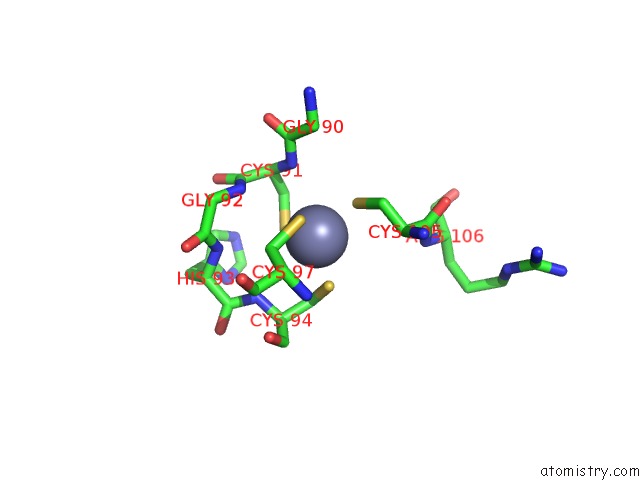
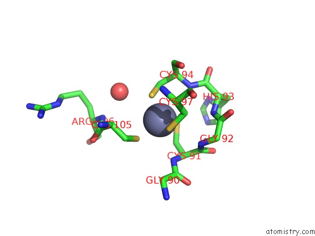
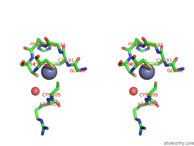
Zinc binding site 1 out of 4 in 4eex
Go back to
Zinc binding site 1 out
of 4 in the Crystal Structure of Lactococcus Lactis Alcohol Dehydrogenase
Mono view
Stereo pair view
Mono view
Stereo pair view
A full contact list of Zinc with other atoms in the Zn binding
site number 1 of Crystal Structure of Lactococcus Lactis Alcohol Dehydrogenase within 5.0Å range:
|
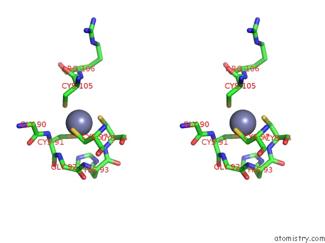
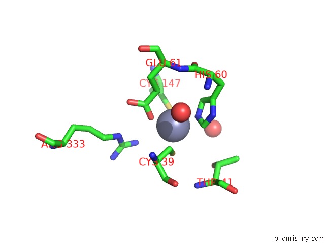
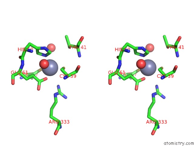
Zinc binding site 2 out of 4 in 4eex
Go back to
Zinc binding site 2 out
of 4 in the Crystal Structure of Lactococcus Lactis Alcohol Dehydrogenase
Mono view
Stereo pair view
Mono view
Stereo pair view
A full contact list of Zinc with other atoms in the Zn binding
site number 2 of Crystal Structure of Lactococcus Lactis Alcohol Dehydrogenase within 5.0Å range:
|
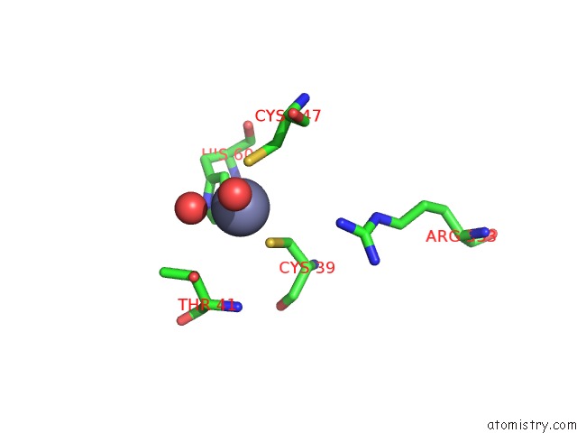
Zinc binding site 3 out of 4 in 4eex
Go back to
Zinc binding site 3 out
of 4 in the Crystal Structure of Lactococcus Lactis Alcohol Dehydrogenase
Mono view
Stereo pair view
Mono view
Stereo pair view
A full contact list of Zinc with other atoms in the Zn binding
site number 3 of Crystal Structure of Lactococcus Lactis Alcohol Dehydrogenase within 5.0Å range:
|
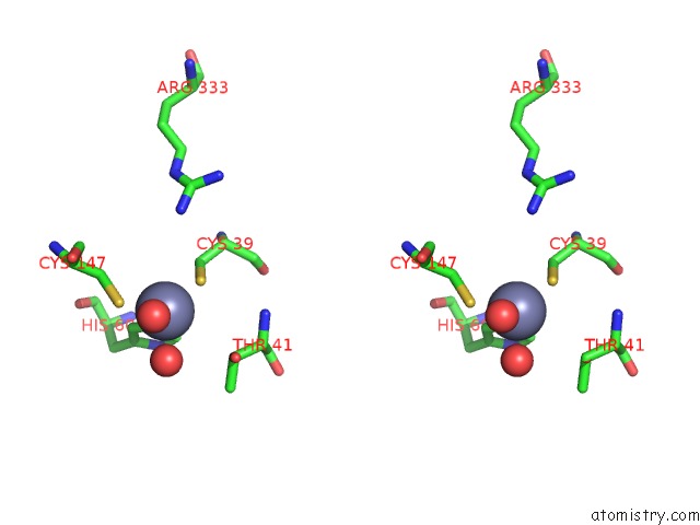
Zinc binding site 4 out of 4 in 4eex
Go back to
Zinc binding site 4 out
of 4 in the Crystal Structure of Lactococcus Lactis Alcohol Dehydrogenase
Mono view
Stereo pair view
Mono view
Stereo pair view
A full contact list of Zinc with other atoms in the Zn binding
site number 4 of Crystal Structure of Lactococcus Lactis Alcohol Dehydrogenase within 5.0Å range:
|
Reference:
X.Liu,
S.Bastian,
C.D.Snow,
E.M.Brustad,
T.E.Saleski,
J.H.Xu,
P.Meinhold,
F.H.Arnold.
Structure-Guided Engineering of Lactococcus Lactis Alcohol Dehydrogenase Lladha For Improved Conversion of Isobutyraldehyde to Isobutanol. J.Biotechnol. V. 164 188 2012.
ISSN: ISSN 0168-1656
PubMed: 22974724
DOI: 10.1016/J.JBIOTEC.2012.08.008
Page generated: Sat Oct 26 21:57:52 2024
ISSN: ISSN 0168-1656
PubMed: 22974724
DOI: 10.1016/J.JBIOTEC.2012.08.008
Last articles
Zn in 9J0NZn in 9J0O
Zn in 9J0P
Zn in 9FJX
Zn in 9EKB
Zn in 9C0F
Zn in 9CAH
Zn in 9CH0
Zn in 9CH3
Zn in 9CH1