Zinc »
PDB 4cz1-4d9q »
4d10 »
Zinc in PDB 4d10: Crystal Structure of the COP9 Signalosome
Protein crystallography data
The structure of Crystal Structure of the COP9 Signalosome, PDB code: 4d10
was solved by
R.D.Bunker,
G.M.Lingaraju,
N.H.Thoma,
with X-Ray Crystallography technique. A brief refinement statistics is given in the table below:
| Resolution Low / High (Å) | 50.87 / 3.80 |
| Space group | P 31 |
| Cell size a, b, c (Å), α, β, γ (°) | 151.615, 151.615, 343.069, 90.00, 90.00, 120.00 |
| R / Rfree (%) | 19.94 / 22.75 |
Zinc Binding Sites:
The binding sites of Zinc atom in the Crystal Structure of the COP9 Signalosome
(pdb code 4d10). This binding sites where shown within
5.0 Angstroms radius around Zinc atom.
In total 2 binding sites of Zinc where determined in the Crystal Structure of the COP9 Signalosome, PDB code: 4d10:
Jump to Zinc binding site number: 1; 2;
In total 2 binding sites of Zinc where determined in the Crystal Structure of the COP9 Signalosome, PDB code: 4d10:
Jump to Zinc binding site number: 1; 2;
Zinc binding site 1 out of 2 in 4d10
Go back to
Zinc binding site 1 out
of 2 in the Crystal Structure of the COP9 Signalosome
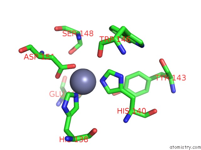
Mono view
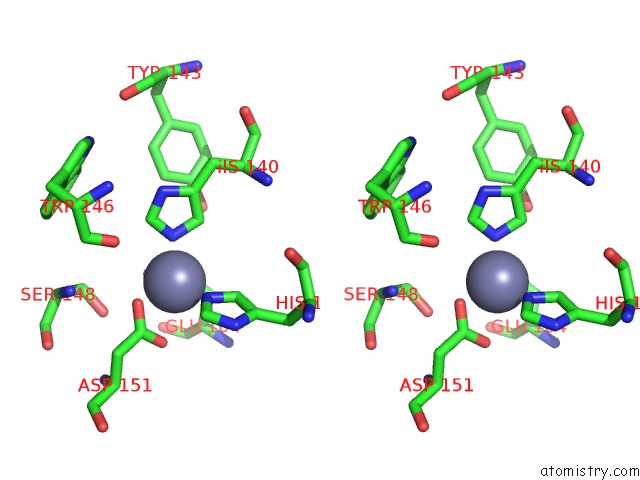
Stereo pair view
Mono view
Stereo pair view
A full contact list of Zinc with other atoms in the Zn binding
site number 1 of Crystal Structure of the COP9 Signalosome within 5.0Å range:
|
Zinc binding site 2 out of 2 in 4d10
Go back to
Zinc binding site 2 out
of 2 in the Crystal Structure of the COP9 Signalosome
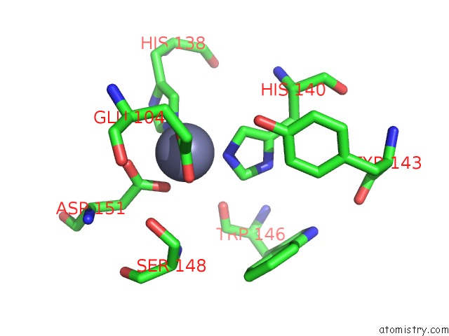
Mono view
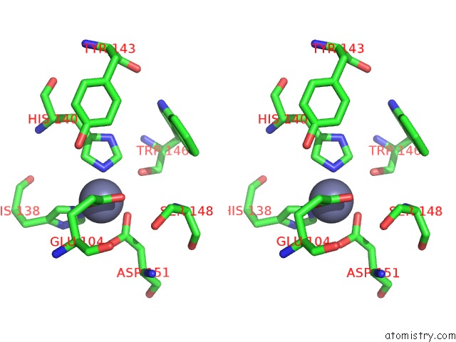
Stereo pair view
Mono view
Stereo pair view
A full contact list of Zinc with other atoms in the Zn binding
site number 2 of Crystal Structure of the COP9 Signalosome within 5.0Å range:
|
Reference:
G.M.Lingaraju,
R.D.Bunker,
S.Cavadini,
D.Hess,
U.Hassiepen,
M.Renatus,
E.S.Fischer,
N.H.Thoma.
Crystal Structure of the Human COP9 Signalosome Nature V. 512 161 2014.
ISSN: ISSN 0028-0836
PubMed: 25043011
DOI: 10.1038/NATURE13566
Page generated: Sat Oct 26 21:12:14 2024
ISSN: ISSN 0028-0836
PubMed: 25043011
DOI: 10.1038/NATURE13566
Last articles
Zn in 9J0NZn in 9J0O
Zn in 9J0P
Zn in 9FJX
Zn in 9EKB
Zn in 9C0F
Zn in 9CAH
Zn in 9CH0
Zn in 9CH3
Zn in 9CH1