Zinc »
PDB 4c6p-4ci3 »
4cc9 »
Zinc in PDB 4cc9: Crystal Structure of Human SAMHD1 (Amino Acid Residues 582- 626) Bound to Vpx Isolated From Sooty Mangabey and Human DCAF1 (Amino Acid Residues 1058-1396)
Protein crystallography data
The structure of Crystal Structure of Human SAMHD1 (Amino Acid Residues 582- 626) Bound to Vpx Isolated From Sooty Mangabey and Human DCAF1 (Amino Acid Residues 1058-1396), PDB code: 4cc9
was solved by
D.Schwefel,
H.C.T.Groom,
V.C.Boucherit,
E.Christodoulou,
P.A.Walker,
J.P.Stoye,
K.N.Bishop,
I.A.Taylor,
with X-Ray Crystallography technique. A brief refinement statistics is given in the table below:
| Resolution Low / High (Å) | 29.227 / 2.47 |
| Space group | P 21 21 21 |
| Cell size a, b, c (Å), α, β, γ (°) | 74.250, 82.881, 115.561, 90.00, 90.00, 90.00 |
| R / Rfree (%) | 17.57 / 21.64 |
Zinc Binding Sites:
The binding sites of Zinc atom in the Crystal Structure of Human SAMHD1 (Amino Acid Residues 582- 626) Bound to Vpx Isolated From Sooty Mangabey and Human DCAF1 (Amino Acid Residues 1058-1396)
(pdb code 4cc9). This binding sites where shown within
5.0 Angstroms radius around Zinc atom.
In total only one binding site of Zinc was determined in the Crystal Structure of Human SAMHD1 (Amino Acid Residues 582- 626) Bound to Vpx Isolated From Sooty Mangabey and Human DCAF1 (Amino Acid Residues 1058-1396), PDB code: 4cc9:
In total only one binding site of Zinc was determined in the Crystal Structure of Human SAMHD1 (Amino Acid Residues 582- 626) Bound to Vpx Isolated From Sooty Mangabey and Human DCAF1 (Amino Acid Residues 1058-1396), PDB code: 4cc9:
Zinc binding site 1 out of 1 in 4cc9
Go back to
Zinc binding site 1 out
of 1 in the Crystal Structure of Human SAMHD1 (Amino Acid Residues 582- 626) Bound to Vpx Isolated From Sooty Mangabey and Human DCAF1 (Amino Acid Residues 1058-1396)
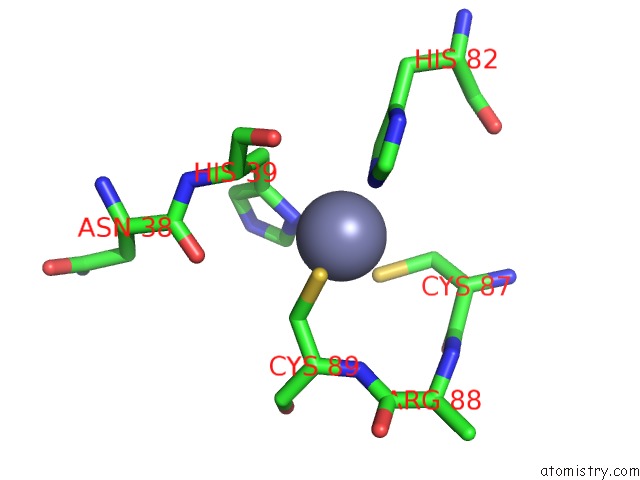
Mono view
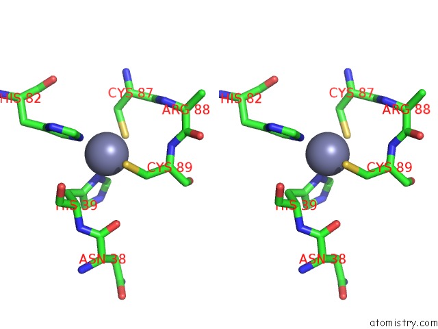
Stereo pair view
Mono view
Stereo pair view
A full contact list of Zinc with other atoms in the Zn binding
site number 1 of Crystal Structure of Human SAMHD1 (Amino Acid Residues 582- 626) Bound to Vpx Isolated From Sooty Mangabey and Human DCAF1 (Amino Acid Residues 1058-1396) within 5.0Å range:
|
Reference:
D.Schwefel,
H.C.T.Groom,
V.C.Boucherit,
E.Christodoulou,
P.A.Walker,
J.P.Stoye,
K.N.Bishop,
I.A.Taylor.
Structural Basis of Lentiviral Subversion of A Cellular Protein Degradation Pathway. Nature V. 505 234 2014.
ISSN: ISSN 0028-0836
PubMed: 24336198
DOI: 10.1038/NATURE12815
Page generated: Sat Oct 26 20:45:33 2024
ISSN: ISSN 0028-0836
PubMed: 24336198
DOI: 10.1038/NATURE12815
Last articles
Zn in 9J0NZn in 9J0O
Zn in 9J0P
Zn in 9FJX
Zn in 9EKB
Zn in 9C0F
Zn in 9CAH
Zn in 9CH0
Zn in 9CH3
Zn in 9CH1