Zinc »
PDB 4c6p-4ci3 »
4ca6 »
Zinc in PDB 4ca6: Human Angiotensin Converting Enzyme N-Domain in Complex with A Phosphinic Tripeptide Fi
Enzymatic activity of Human Angiotensin Converting Enzyme N-Domain in Complex with A Phosphinic Tripeptide Fi
All present enzymatic activity of Human Angiotensin Converting Enzyme N-Domain in Complex with A Phosphinic Tripeptide Fi:
3.4.15.1;
3.4.15.1;
Protein crystallography data
The structure of Human Angiotensin Converting Enzyme N-Domain in Complex with A Phosphinic Tripeptide Fi, PDB code: 4ca6
was solved by
G.Masuyer,
M.Akif,
B.Czarny,
F.Beau,
S.L.U.Schwager,
E.D.Sturrock,
R.E.Isaac,
V.Dive,
K.R.Acharya,
with X-Ray Crystallography technique. A brief refinement statistics is given in the table below:
| Resolution Low / High (Å) | 29.74 / 1.91 |
| Space group | P 1 |
| Cell size a, b, c (Å), α, β, γ (°) | 72.925, 76.643, 82.545, 88.62, 64.22, 75.58 |
| R / Rfree (%) | 18.675 / 22.296 |
Other elements in 4ca6:
The structure of Human Angiotensin Converting Enzyme N-Domain in Complex with A Phosphinic Tripeptide Fi also contains other interesting chemical elements:
| Chlorine | (Cl) | 2 atoms |
Zinc Binding Sites:
The binding sites of Zinc atom in the Human Angiotensin Converting Enzyme N-Domain in Complex with A Phosphinic Tripeptide Fi
(pdb code 4ca6). This binding sites where shown within
5.0 Angstroms radius around Zinc atom.
In total 2 binding sites of Zinc where determined in the Human Angiotensin Converting Enzyme N-Domain in Complex with A Phosphinic Tripeptide Fi, PDB code: 4ca6:
Jump to Zinc binding site number: 1; 2;
In total 2 binding sites of Zinc where determined in the Human Angiotensin Converting Enzyme N-Domain in Complex with A Phosphinic Tripeptide Fi, PDB code: 4ca6:
Jump to Zinc binding site number: 1; 2;
Zinc binding site 1 out of 2 in 4ca6
Go back to
Zinc binding site 1 out
of 2 in the Human Angiotensin Converting Enzyme N-Domain in Complex with A Phosphinic Tripeptide Fi
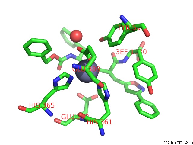
Mono view
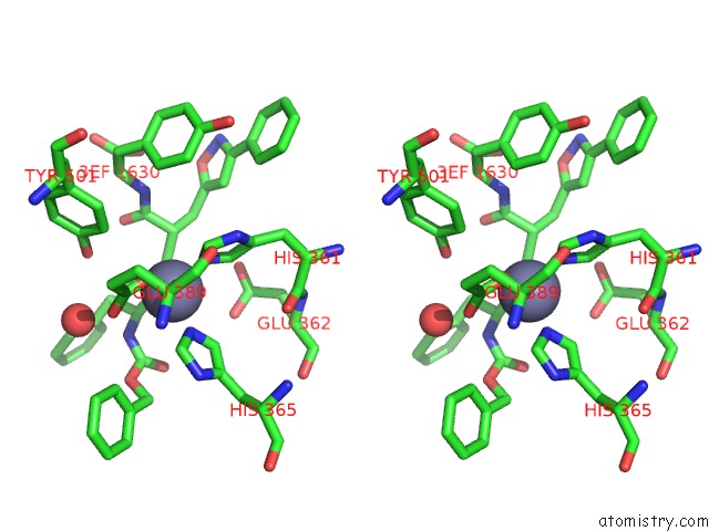
Stereo pair view
Mono view
Stereo pair view
A full contact list of Zinc with other atoms in the Zn binding
site number 1 of Human Angiotensin Converting Enzyme N-Domain in Complex with A Phosphinic Tripeptide Fi within 5.0Å range:
|
Zinc binding site 2 out of 2 in 4ca6
Go back to
Zinc binding site 2 out
of 2 in the Human Angiotensin Converting Enzyme N-Domain in Complex with A Phosphinic Tripeptide Fi
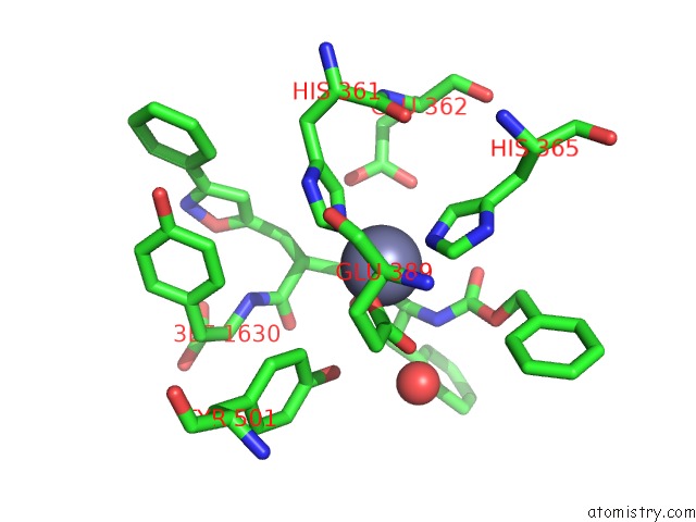
Mono view
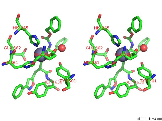
Stereo pair view
Mono view
Stereo pair view
A full contact list of Zinc with other atoms in the Zn binding
site number 2 of Human Angiotensin Converting Enzyme N-Domain in Complex with A Phosphinic Tripeptide Fi within 5.0Å range:
|
Reference:
G.Masuyer,
M.Akif,
B.Czarny,
F.Beau,
S.L.Schwager,
E.D.Sturrock,
R.E.Isaac,
V.Dive,
K.R.Acharya.
Crystal Structures of Highly Specific Phosphinic Tripeptide Enantiomers in Complex with the Angiotensin-I Converting Enzyme. Febs J. V. 281 943 2014.
ISSN: ISSN 1742-464X
PubMed: 24289879
DOI: 10.1111/FEBS.12660
Page generated: Sat Oct 26 20:42:33 2024
ISSN: ISSN 1742-464X
PubMed: 24289879
DOI: 10.1111/FEBS.12660
Last articles
Zn in 9J0NZn in 9J0O
Zn in 9J0P
Zn in 9FJX
Zn in 9EKB
Zn in 9C0F
Zn in 9CAH
Zn in 9CH0
Zn in 9CH3
Zn in 9CH1