Zinc »
PDB 4c1d-4c6o »
4c4a »
Zinc in PDB 4c4a: Crystal Structure of Mouse Protein Arginine Methyltransferase 7 in Complex with Sah
Enzymatic activity of Crystal Structure of Mouse Protein Arginine Methyltransferase 7 in Complex with Sah
All present enzymatic activity of Crystal Structure of Mouse Protein Arginine Methyltransferase 7 in Complex with Sah:
2.1.1.125; 2.1.1.126;
2.1.1.125; 2.1.1.126;
Protein crystallography data
The structure of Crystal Structure of Mouse Protein Arginine Methyltransferase 7 in Complex with Sah, PDB code: 4c4a
was solved by
V.Cura,
N.Troffer-Charlier,
L.Bonnefond,
J.M.Wurtz,
J.Cavarelli,
with X-Ray Crystallography technique. A brief refinement statistics is given in the table below:
| Resolution Low / High (Å) | 29.353 / 1.70 |
| Space group | P 43 21 2 |
| Cell size a, b, c (Å), α, β, γ (°) | 97.322, 97.322, 168.754, 90.00, 90.00, 90.00 |
| R / Rfree (%) | 15.97 / 18.97 |
Zinc Binding Sites:
The binding sites of Zinc atom in the Crystal Structure of Mouse Protein Arginine Methyltransferase 7 in Complex with Sah
(pdb code 4c4a). This binding sites where shown within
5.0 Angstroms radius around Zinc atom.
In total only one binding site of Zinc was determined in the Crystal Structure of Mouse Protein Arginine Methyltransferase 7 in Complex with Sah, PDB code: 4c4a:
In total only one binding site of Zinc was determined in the Crystal Structure of Mouse Protein Arginine Methyltransferase 7 in Complex with Sah, PDB code: 4c4a:
Zinc binding site 1 out of 1 in 4c4a
Go back to
Zinc binding site 1 out
of 1 in the Crystal Structure of Mouse Protein Arginine Methyltransferase 7 in Complex with Sah
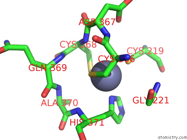
Mono view
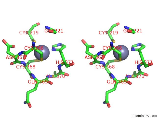
Stereo pair view
Mono view
Stereo pair view
A full contact list of Zinc with other atoms in the Zn binding
site number 1 of Crystal Structure of Mouse Protein Arginine Methyltransferase 7 in Complex with Sah within 5.0Å range:
|
Reference:
V.Cura,
N.Troffer-Charlier,
J.M.Wurtz,
L.Bonnefond,
J.Cavarelli.
Structural Insight Into Arginine Methylation By the Mouse Protein Arginine Methyltransferase 7: A Zinc Finger Freezes the Mimic of the Dimeric State Into A Single Active Site. Acta Crystallogr.,Sect.D V. 70 2401 2014.
ISSN: ISSN 0907-4449
PubMed: 25195753
DOI: 10.1107/S1399004714014278
Page generated: Sat Oct 26 20:23:37 2024
ISSN: ISSN 0907-4449
PubMed: 25195753
DOI: 10.1107/S1399004714014278
Last articles
Zn in 9J0NZn in 9J0O
Zn in 9J0P
Zn in 9FJX
Zn in 9EKB
Zn in 9C0F
Zn in 9CAH
Zn in 9CH0
Zn in 9CH3
Zn in 9CH1