Zinc »
PDB 4bjb-4bud »
4bjc »
Zinc in PDB 4bjc: Crystal Structure of Human Tankyrase 2 in Complex with Rucaparib
Enzymatic activity of Crystal Structure of Human Tankyrase 2 in Complex with Rucaparib
All present enzymatic activity of Crystal Structure of Human Tankyrase 2 in Complex with Rucaparib:
2.4.2.30;
2.4.2.30;
Protein crystallography data
The structure of Crystal Structure of Human Tankyrase 2 in Complex with Rucaparib, PDB code: 4bjc
was solved by
T.Haikarainen,
M.Narwal,
L.Lehtio,
with X-Ray Crystallography technique. A brief refinement statistics is given in the table below:
| Resolution Low / High (Å) | 29.20 / 2.20 |
| Space group | P 41 21 2 |
| Cell size a, b, c (Å), α, β, γ (°) | 65.300, 65.300, 122.910, 90.00, 90.00, 90.00 |
| R / Rfree (%) | 19.458 / 24.718 |
Other elements in 4bjc:
The structure of Crystal Structure of Human Tankyrase 2 in Complex with Rucaparib also contains other interesting chemical elements:
| Fluorine | (F) | 1 atom |
Zinc Binding Sites:
The binding sites of Zinc atom in the Crystal Structure of Human Tankyrase 2 in Complex with Rucaparib
(pdb code 4bjc). This binding sites where shown within
5.0 Angstroms radius around Zinc atom.
In total only one binding site of Zinc was determined in the Crystal Structure of Human Tankyrase 2 in Complex with Rucaparib, PDB code: 4bjc:
In total only one binding site of Zinc was determined in the Crystal Structure of Human Tankyrase 2 in Complex with Rucaparib, PDB code: 4bjc:
Zinc binding site 1 out of 1 in 4bjc
Go back to
Zinc binding site 1 out
of 1 in the Crystal Structure of Human Tankyrase 2 in Complex with Rucaparib
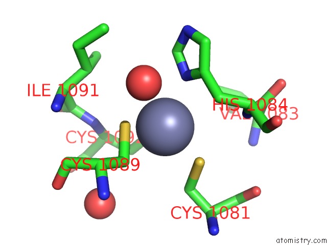
Mono view
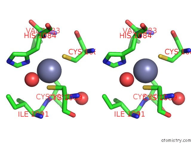
Stereo pair view
Mono view
Stereo pair view
A full contact list of Zinc with other atoms in the Zn binding
site number 1 of Crystal Structure of Human Tankyrase 2 in Complex with Rucaparib within 5.0Å range:
|
Reference:
T.Haikarainen,
M.Narwal,
P.Joensuu,
L.Lehtio.
Evaluation and Structural Basis For the Inhibition of Tankyrases By Parp Inhibitors Acs Med.Chem.Lett. V. 5 18 2014.
ISSN: ISSN 1948-5875
PubMed: 24900770
DOI: 10.1021/ML400292S
Page generated: Sat Oct 26 19:49:38 2024
ISSN: ISSN 1948-5875
PubMed: 24900770
DOI: 10.1021/ML400292S
Last articles
Zn in 9J0NZn in 9J0O
Zn in 9J0P
Zn in 9FJX
Zn in 9EKB
Zn in 9C0F
Zn in 9CAH
Zn in 9CH0
Zn in 9CH3
Zn in 9CH1