Zinc »
PDB 4ask-4b3t »
4av1 »
Zinc in PDB 4av1: Crystal Structure of the Human Parp-1 Dna Binding Domain in Complex with Dna
Enzymatic activity of Crystal Structure of the Human Parp-1 Dna Binding Domain in Complex with Dna
All present enzymatic activity of Crystal Structure of the Human Parp-1 Dna Binding Domain in Complex with Dna:
2.4.2.30;
2.4.2.30;
Protein crystallography data
The structure of Crystal Structure of the Human Parp-1 Dna Binding Domain in Complex with Dna, PDB code: 4av1
was solved by
A.A.E.Ali,
G.Timinszky,
R.Arribas-Bosacoma,
M.Kozlowski,
P.O.Hassa,
M.Hassler,
A.G.Ladurner,
L.H.Pearl,
A.W.Oliver,
with X-Ray Crystallography technique. A brief refinement statistics is given in the table below:
| Resolution Low / High (Å) | 44.353 / 3.10 |
| Space group | C 1 2 1 |
| Cell size a, b, c (Å), α, β, γ (°) | 163.978, 59.504, 61.582, 90.00, 101.18, 90.00 |
| R / Rfree (%) | 22.95 / 24.87 |
Zinc Binding Sites:
The binding sites of Zinc atom in the Crystal Structure of the Human Parp-1 Dna Binding Domain in Complex with Dna
(pdb code 4av1). This binding sites where shown within
5.0 Angstroms radius around Zinc atom.
In total 4 binding sites of Zinc where determined in the Crystal Structure of the Human Parp-1 Dna Binding Domain in Complex with Dna, PDB code: 4av1:
Jump to Zinc binding site number: 1; 2; 3; 4;
In total 4 binding sites of Zinc where determined in the Crystal Structure of the Human Parp-1 Dna Binding Domain in Complex with Dna, PDB code: 4av1:
Jump to Zinc binding site number: 1; 2; 3; 4;
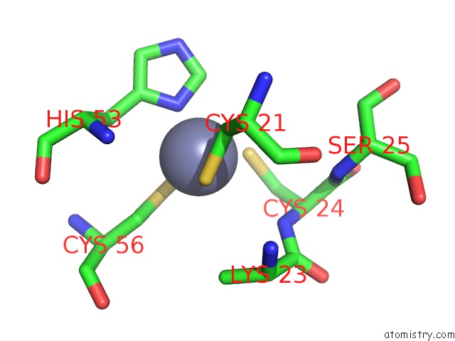
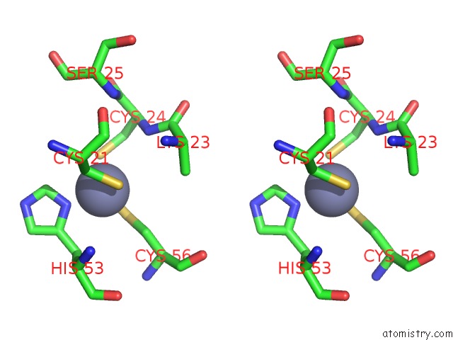
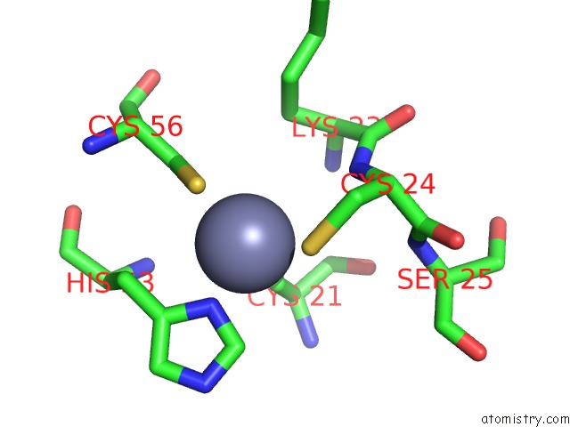
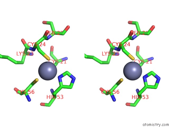
Zinc binding site 1 out of 4 in 4av1
Go back to
Zinc binding site 1 out
of 4 in the Crystal Structure of the Human Parp-1 Dna Binding Domain in Complex with Dna
Mono view
Stereo pair view
Mono view
Stereo pair view
A full contact list of Zinc with other atoms in the Zn binding
site number 1 of Crystal Structure of the Human Parp-1 Dna Binding Domain in Complex with Dna within 5.0Å range:
|
Zinc binding site 2 out of 4 in 4av1
Go back to
Zinc binding site 2 out
of 4 in the Crystal Structure of the Human Parp-1 Dna Binding Domain in Complex with Dna
Mono view
Stereo pair view
Mono view
Stereo pair view
A full contact list of Zinc with other atoms in the Zn binding
site number 2 of Crystal Structure of the Human Parp-1 Dna Binding Domain in Complex with Dna within 5.0Å range:
|
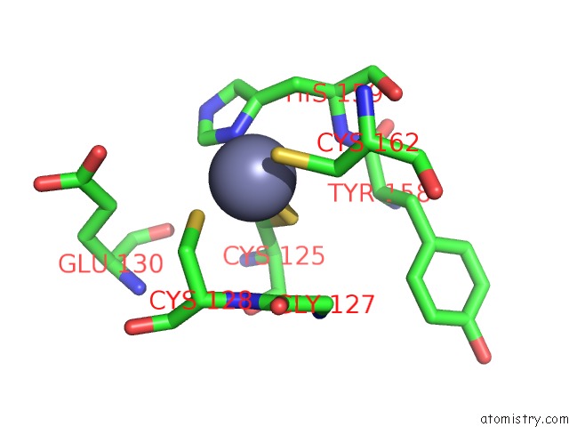
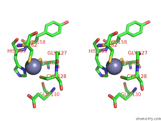
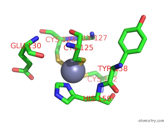
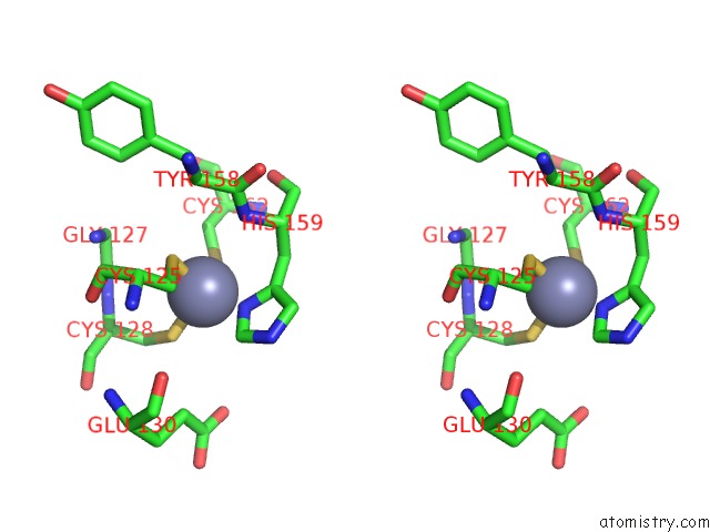
Zinc binding site 3 out of 4 in 4av1
Go back to
Zinc binding site 3 out
of 4 in the Crystal Structure of the Human Parp-1 Dna Binding Domain in Complex with Dna
Mono view
Stereo pair view
Mono view
Stereo pair view
A full contact list of Zinc with other atoms in the Zn binding
site number 3 of Crystal Structure of the Human Parp-1 Dna Binding Domain in Complex with Dna within 5.0Å range:
|
Zinc binding site 4 out of 4 in 4av1
Go back to
Zinc binding site 4 out
of 4 in the Crystal Structure of the Human Parp-1 Dna Binding Domain in Complex with Dna
Mono view
Stereo pair view
Mono view
Stereo pair view
A full contact list of Zinc with other atoms in the Zn binding
site number 4 of Crystal Structure of the Human Parp-1 Dna Binding Domain in Complex with Dna within 5.0Å range:
|
Reference:
A.A.E.Ali,
G.Timinszky,
R.Arribas-Bosacoma,
M.Kozlowski,
P.O.Hassa,
M.Hassler,
A.G.Ladurner,
L.H.Pearl,
A.W.Oliver.
The Zinc-Finger Domains of PARP1 Cooperate to Recognise Dna Strand-Breaks Nat.Struct.Mol.Biol. V. 19 685 2012.
ISSN: ISSN 1072-8368
PubMed: 22683995
DOI: 10.1038/NSMB.2335
Page generated: Sat Oct 26 19:23:24 2024
ISSN: ISSN 1072-8368
PubMed: 22683995
DOI: 10.1038/NSMB.2335
Last articles
Zn in 9J0NZn in 9J0O
Zn in 9J0P
Zn in 9FJX
Zn in 9EKB
Zn in 9C0F
Zn in 9CAH
Zn in 9CH0
Zn in 9CH3
Zn in 9CH1