Zinc »
PDB 3x17-3zr9 »
3zqr »
Zinc in PDB 3zqr: NMEPHEB25 Insulin Analogue Crystal Structure
Protein crystallography data
The structure of NMEPHEB25 Insulin Analogue Crystal Structure, PDB code: 3zqr
was solved by
E.Antolikova,
L.Zakova,
J.P.Turkenburg,
C.J.Watson,
I.Hanclova,
M.Sanda,
A.Cooper,
T.Kraus,
A.M.Brzozowski,
J.A.Jiracek,
with X-Ray Crystallography technique. A brief refinement statistics is given in the table below:
| Resolution Low / High (Å) | 54.29 / 1.90 |
| Space group | P 1 21 1 |
| Cell size a, b, c (Å), α, β, γ (°) | 46.600, 61.837, 58.224, 90.00, 111.19, 90.00 |
| R / Rfree (%) | 22.3 / 28.1 |
Other elements in 3zqr:
The structure of NMEPHEB25 Insulin Analogue Crystal Structure also contains other interesting chemical elements:
| Chlorine | (Cl) | 2 atoms |
Zinc Binding Sites:
The binding sites of Zinc atom in the NMEPHEB25 Insulin Analogue Crystal Structure
(pdb code 3zqr). This binding sites where shown within
5.0 Angstroms radius around Zinc atom.
In total 2 binding sites of Zinc where determined in the NMEPHEB25 Insulin Analogue Crystal Structure, PDB code: 3zqr:
Jump to Zinc binding site number: 1; 2;
In total 2 binding sites of Zinc where determined in the NMEPHEB25 Insulin Analogue Crystal Structure, PDB code: 3zqr:
Jump to Zinc binding site number: 1; 2;
Zinc binding site 1 out of 2 in 3zqr
Go back to
Zinc binding site 1 out
of 2 in the NMEPHEB25 Insulin Analogue Crystal Structure
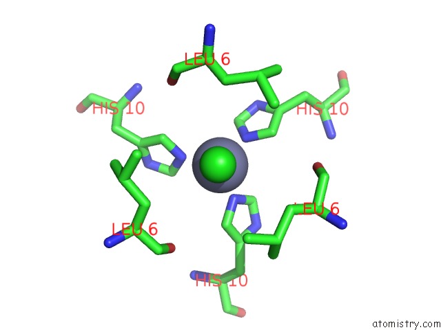
Mono view
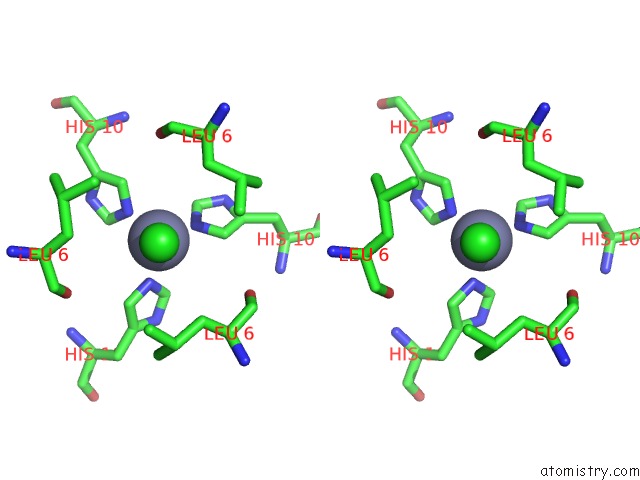
Stereo pair view
Mono view
Stereo pair view
A full contact list of Zinc with other atoms in the Zn binding
site number 1 of NMEPHEB25 Insulin Analogue Crystal Structure within 5.0Å range:
|
Zinc binding site 2 out of 2 in 3zqr
Go back to
Zinc binding site 2 out
of 2 in the NMEPHEB25 Insulin Analogue Crystal Structure
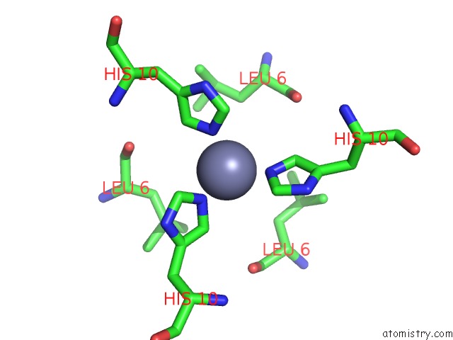
Mono view
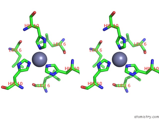
Stereo pair view
Mono view
Stereo pair view
A full contact list of Zinc with other atoms in the Zn binding
site number 2 of NMEPHEB25 Insulin Analogue Crystal Structure within 5.0Å range:
|
Reference:
E.Antolikova,
L.Zakova,
J.P.Turkenburg,
C.J.Watson,
I.Hanclova,
M.Sanda,
A.Cooper,
T.Kraus,
A.M.Brzozowski,
J.A.Jiracek.
Non-Equivalent Role of Inter- and Intramolecular Hydrogen Bonds in the Insulin Dimer Interface. J.Biol.Chem. V. 286 36968 2011.
ISSN: ISSN 0021-9258
PubMed: 21880708
DOI: 10.1074/JBC.M111.265249
Page generated: Sat Oct 26 18:34:21 2024
ISSN: ISSN 0021-9258
PubMed: 21880708
DOI: 10.1074/JBC.M111.265249
Last articles
Zn in 9J0NZn in 9J0O
Zn in 9J0P
Zn in 9FJX
Zn in 9EKB
Zn in 9C0F
Zn in 9CAH
Zn in 9CH0
Zn in 9CH3
Zn in 9CH1