Zinc »
PDB 3uk4-3v38 »
3umi »
Zinc in PDB 3umi: X-Ray Structure of the E2 Domain of the Human Amyloid Precursor Protein (App) in Complex with Zinc
Protein crystallography data
The structure of X-Ray Structure of the E2 Domain of the Human Amyloid Precursor Protein (App) in Complex with Zinc, PDB code: 3umi
was solved by
S.O.Dahms,
I.Konnig,
D.Roeser,
K.H.Guhrs,
M.E.Than,
with X-Ray Crystallography technique. A brief refinement statistics is given in the table below:
| Resolution Low / High (Å) | 27.44 / 2.40 |
| Space group | P 43 |
| Cell size a, b, c (Å), α, β, γ (°) | 39.757, 39.757, 126.425, 90.00, 90.00, 90.00 |
| R / Rfree (%) | 20.4 / 24.5 |
Other elements in 3umi:
The structure of X-Ray Structure of the E2 Domain of the Human Amyloid Precursor Protein (App) in Complex with Zinc also contains other interesting chemical elements:
| Cadmium | (Cd) | 9 atoms |
Zinc Binding Sites:
The binding sites of Zinc atom in the X-Ray Structure of the E2 Domain of the Human Amyloid Precursor Protein (App) in Complex with Zinc
(pdb code 3umi). This binding sites where shown within
5.0 Angstroms radius around Zinc atom.
In total only one binding site of Zinc was determined in the X-Ray Structure of the E2 Domain of the Human Amyloid Precursor Protein (App) in Complex with Zinc, PDB code: 3umi:
In total only one binding site of Zinc was determined in the X-Ray Structure of the E2 Domain of the Human Amyloid Precursor Protein (App) in Complex with Zinc, PDB code: 3umi:
Zinc binding site 1 out of 1 in 3umi
Go back to
Zinc binding site 1 out
of 1 in the X-Ray Structure of the E2 Domain of the Human Amyloid Precursor Protein (App) in Complex with Zinc
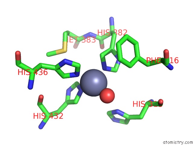
Mono view
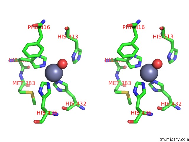
Stereo pair view
Mono view
Stereo pair view
A full contact list of Zinc with other atoms in the Zn binding
site number 1 of X-Ray Structure of the E2 Domain of the Human Amyloid Precursor Protein (App) in Complex with Zinc within 5.0Å range:
|
Reference:
S.O.Dahms,
I.Konnig,
D.Roeser,
K.H.Guhrs,
M.C.Mayer,
D.Kaden,
G.Multhaup,
M.E.Than.
Metal Binding Dictates Conformation and Function of the Amyloid Precursor Protein (App) E2 Domain. J.Mol.Biol. V. 416 438 2012.
ISSN: ISSN 0022-2836
PubMed: 22245578
DOI: 10.1016/J.JMB.2011.12.057
Page generated: Sat Oct 26 17:24:22 2024
ISSN: ISSN 0022-2836
PubMed: 22245578
DOI: 10.1016/J.JMB.2011.12.057
Last articles
Zn in 9J0NZn in 9J0O
Zn in 9J0P
Zn in 9FJX
Zn in 9EKB
Zn in 9C0F
Zn in 9CAH
Zn in 9CH0
Zn in 9CH3
Zn in 9CH1