Zinc »
PDB 3suf-3t6p »
3t0c »
Zinc in PDB 3t0c: Crystal Structure of Streptococcus Mutans Mete Complexed with Zinc
Enzymatic activity of Crystal Structure of Streptococcus Mutans Mete Complexed with Zinc
All present enzymatic activity of Crystal Structure of Streptococcus Mutans Mete Complexed with Zinc:
2.1.1.14;
2.1.1.14;
Protein crystallography data
The structure of Crystal Structure of Streptococcus Mutans Mete Complexed with Zinc, PDB code: 3t0c
was solved by
T.M.Fu,
Y.H.Liang,
X.D.Su,
with X-Ray Crystallography technique. A brief refinement statistics is given in the table below:
| Resolution Low / High (Å) | 42.07 / 2.19 |
| Space group | P 1 21 1 |
| Cell size a, b, c (Å), α, β, γ (°) | 52.845, 99.477, 77.883, 90.00, 94.55, 90.00 |
| R / Rfree (%) | 20.9 / 25.6 |
Zinc Binding Sites:
The binding sites of Zinc atom in the Crystal Structure of Streptococcus Mutans Mete Complexed with Zinc
(pdb code 3t0c). This binding sites where shown within
5.0 Angstroms radius around Zinc atom.
In total only one binding site of Zinc was determined in the Crystal Structure of Streptococcus Mutans Mete Complexed with Zinc, PDB code: 3t0c:
In total only one binding site of Zinc was determined in the Crystal Structure of Streptococcus Mutans Mete Complexed with Zinc, PDB code: 3t0c:
Zinc binding site 1 out of 1 in 3t0c
Go back to
Zinc binding site 1 out
of 1 in the Crystal Structure of Streptococcus Mutans Mete Complexed with Zinc
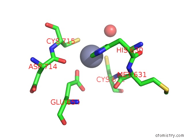
Mono view
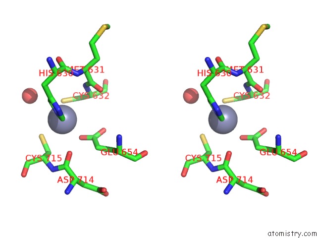
Stereo pair view
Mono view
Stereo pair view
A full contact list of Zinc with other atoms in the Zn binding
site number 1 of Crystal Structure of Streptococcus Mutans Mete Complexed with Zinc within 5.0Å range:
|
Reference:
T.M.Fu,
J.Almqvist,
Y.H.Liang,
L.Li,
Y.Huang,
X.D.Su.
Crystal Structures of Cobalamin-Independent Methionine Synthase (Mete) From Streptococcus Mutans: A Dynamic Zinc-Inversion Model J.Mol.Biol. V. 412 688 2011.
ISSN: ISSN 0022-2836
PubMed: 21840320
DOI: 10.1016/J.JMB.2011.08.005
Page generated: Sat Oct 26 16:15:15 2024
ISSN: ISSN 0022-2836
PubMed: 21840320
DOI: 10.1016/J.JMB.2011.08.005
Last articles
Zn in 9J0NZn in 9J0O
Zn in 9J0P
Zn in 9FJX
Zn in 9EKB
Zn in 9C0F
Zn in 9CAH
Zn in 9CH0
Zn in 9CH3
Zn in 9CH1