Zinc »
PDB 3sck-3sjd »
3si0 »
Zinc in PDB 3si0: Structure of Glycosylated Human Glutaminyl Cyclase
Enzymatic activity of Structure of Glycosylated Human Glutaminyl Cyclase
All present enzymatic activity of Structure of Glycosylated Human Glutaminyl Cyclase:
2.3.2.5;
2.3.2.5;
Protein crystallography data
The structure of Structure of Glycosylated Human Glutaminyl Cyclase, PDB code: 3si0
was solved by
C.Parthier,
D.Carrillo,
M.T.Stubbs,
with X-Ray Crystallography technique. A brief refinement statistics is given in the table below:
| Resolution Low / High (Å) | 11.98 / 2.10 |
| Space group | C 1 2 1 |
| Cell size a, b, c (Å), α, β, γ (°) | 82.408, 63.688, 77.159, 90.00, 105.76, 90.00 |
| R / Rfree (%) | 20.4 / 26.4 |
Other elements in 3si0:
The structure of Structure of Glycosylated Human Glutaminyl Cyclase also contains other interesting chemical elements:
| Chlorine | (Cl) | 1 atom |
Zinc Binding Sites:
The binding sites of Zinc atom in the Structure of Glycosylated Human Glutaminyl Cyclase
(pdb code 3si0). This binding sites where shown within
5.0 Angstroms radius around Zinc atom.
In total only one binding site of Zinc was determined in the Structure of Glycosylated Human Glutaminyl Cyclase, PDB code: 3si0:
In total only one binding site of Zinc was determined in the Structure of Glycosylated Human Glutaminyl Cyclase, PDB code: 3si0:
Zinc binding site 1 out of 1 in 3si0
Go back to
Zinc binding site 1 out
of 1 in the Structure of Glycosylated Human Glutaminyl Cyclase
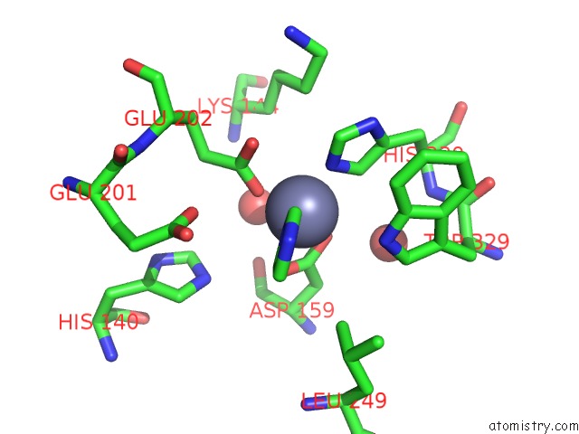
Mono view
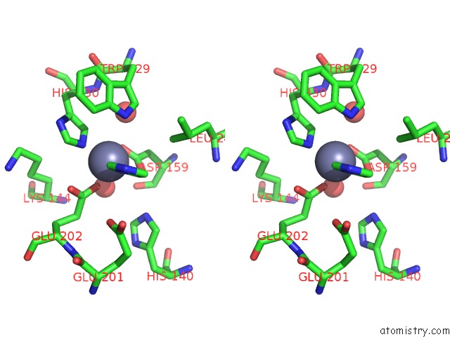
Stereo pair view
Mono view
Stereo pair view
A full contact list of Zinc with other atoms in the Zn binding
site number 1 of Structure of Glycosylated Human Glutaminyl Cyclase within 5.0Å range:
|
Reference:
D.Ruiz-Carrillo,
B.Koch,
C.Parthier,
M.Wermann,
T.Dambe,
M.Buchholz,
H.H.Ludwig,
U.Heiser,
J.U.Rahfeld,
M.T.Stubbs,
S.Schilling,
H.U.Demuth.
Structures of Glycosylated Mammalian Glutaminyl Cyclases Reveal Conformational Variability Near the Active Center. Biochemistry V. 50 6280 2011.
ISSN: ISSN 0006-2960
PubMed: 21671571
DOI: 10.1021/BI200249H
Page generated: Sat Oct 26 15:46:18 2024
ISSN: ISSN 0006-2960
PubMed: 21671571
DOI: 10.1021/BI200249H
Last articles
Zn in 9J0NZn in 9J0O
Zn in 9J0P
Zn in 9FJX
Zn in 9EKB
Zn in 9C0F
Zn in 9CAH
Zn in 9CH0
Zn in 9CH3
Zn in 9CH1