Zinc »
PDB 3sck-3sjd »
3shb »
Zinc in PDB 3shb: Crystal Structure of Phd Domain of UHRF1
Protein crystallography data
The structure of Crystal Structure of Phd Domain of UHRF1, PDB code: 3shb
was solved by
L.Hu,
Z.Li,
P.Wang,
Y.Lin,
Y.Xu,
with X-Ray Crystallography technique. A brief refinement statistics is given in the table below:
| Resolution Low / High (Å) | 31.70 / 1.80 |
| Space group | P 61 2 2 |
| Cell size a, b, c (Å), α, β, γ (°) | 36.609, 36.609, 219.765, 90.00, 90.00, 120.00 |
| R / Rfree (%) | 21 / 22.8 |
Zinc Binding Sites:
The binding sites of Zinc atom in the Crystal Structure of Phd Domain of UHRF1
(pdb code 3shb). This binding sites where shown within
5.0 Angstroms radius around Zinc atom.
In total 4 binding sites of Zinc where determined in the Crystal Structure of Phd Domain of UHRF1, PDB code: 3shb:
Jump to Zinc binding site number: 1; 2; 3; 4;
In total 4 binding sites of Zinc where determined in the Crystal Structure of Phd Domain of UHRF1, PDB code: 3shb:
Jump to Zinc binding site number: 1; 2; 3; 4;
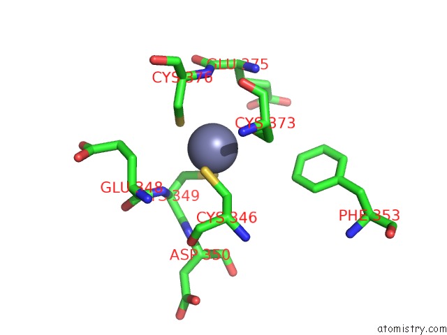
Zinc binding site 1 out of 4 in 3shb
Go back to
Zinc binding site 1 out
of 4 in the Crystal Structure of Phd Domain of UHRF1
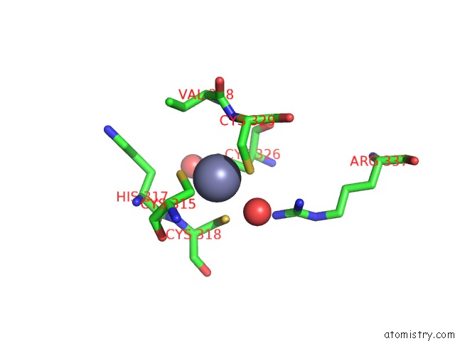
Mono view
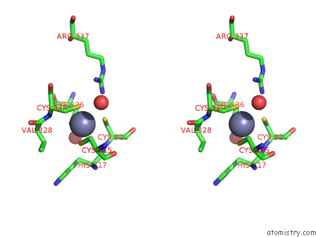
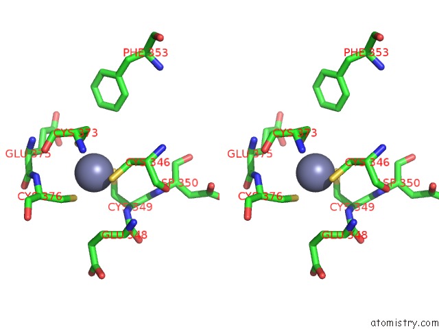
Stereo pair view
Mono view
Stereo pair view
A full contact list of Zinc with other atoms in the Zn binding
site number 1 of Crystal Structure of Phd Domain of UHRF1 within 5.0Å range:
|
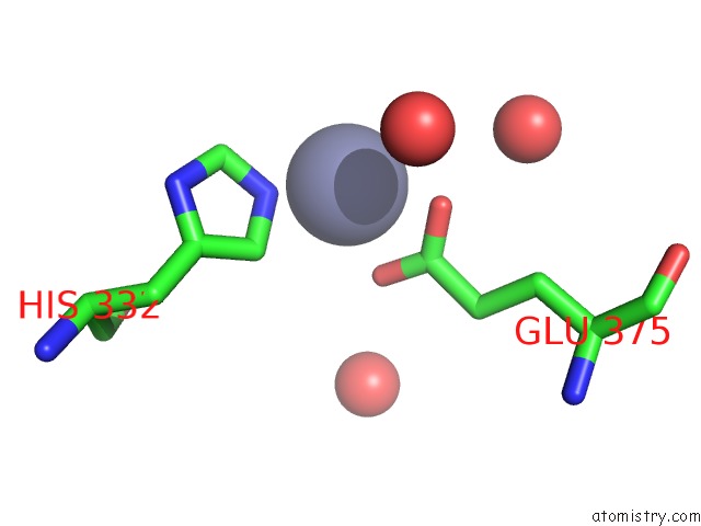
Zinc binding site 2 out of 4 in 3shb
Go back to
Zinc binding site 2 out
of 4 in the Crystal Structure of Phd Domain of UHRF1
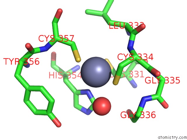
Mono view
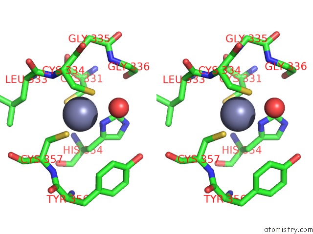
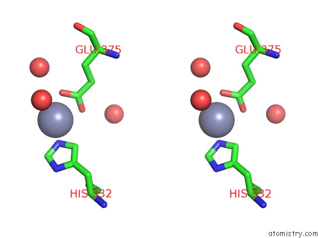
Stereo pair view
Mono view
Stereo pair view
A full contact list of Zinc with other atoms in the Zn binding
site number 2 of Crystal Structure of Phd Domain of UHRF1 within 5.0Å range:
|
Zinc binding site 3 out of 4 in 3shb
Go back to
Zinc binding site 3 out
of 4 in the Crystal Structure of Phd Domain of UHRF1
Mono view
Stereo pair view
Mono view
Stereo pair view
A full contact list of Zinc with other atoms in the Zn binding
site number 3 of Crystal Structure of Phd Domain of UHRF1 within 5.0Å range:
|
Zinc binding site 4 out of 4 in 3shb
Go back to
Zinc binding site 4 out
of 4 in the Crystal Structure of Phd Domain of UHRF1
Mono view
Stereo pair view
Mono view
Stereo pair view
A full contact list of Zinc with other atoms in the Zn binding
site number 4 of Crystal Structure of Phd Domain of UHRF1 within 5.0Å range:
|
Reference:
L.Hu,
Z.Li,
P.Wang,
Y.Lin,
Y.Xu.
Crystal Structure of Phd Domain of UHRF1 and Insights Into Recognition of Unmodified Histone H3 Arginine Residue 2. Cell Res. 2011.
ISSN: ISSN 1001-0602
PubMed: 21808300
DOI: 10.1038/CR.2011.124
Page generated: Sat Oct 26 15:45:28 2024
ISSN: ISSN 1001-0602
PubMed: 21808300
DOI: 10.1038/CR.2011.124
Last articles
Zn in 9J0NZn in 9J0O
Zn in 9J0P
Zn in 9FJX
Zn in 9EKB
Zn in 9C0F
Zn in 9CAH
Zn in 9CH0
Zn in 9CH3
Zn in 9CH1