Zinc »
PDB 3sck-3sjd »
3se6 »
Zinc in PDB 3se6: Crystal Structure of the Human Endoplasmic Reticulum Aminopeptidase 2
Protein crystallography data
The structure of Crystal Structure of the Human Endoplasmic Reticulum Aminopeptidase 2, PDB code: 3se6
was solved by
J.R.Birtley,
E.Saridakis,
E.Stratikos,
I.M.Mavridis,
with X-Ray Crystallography technique. A brief refinement statistics is given in the table below:
| Resolution Low / High (Å) | 11.00 / 3.08 |
| Space group | P 1 21 1 |
| Cell size a, b, c (Å), α, β, γ (°) | 74.579, 134.362, 128.009, 90.00, 90.71, 90.00 |
| R / Rfree (%) | 21.2 / 27.7 |
Zinc Binding Sites:
The binding sites of Zinc atom in the Crystal Structure of the Human Endoplasmic Reticulum Aminopeptidase 2
(pdb code 3se6). This binding sites where shown within
5.0 Angstroms radius around Zinc atom.
In total 2 binding sites of Zinc where determined in the Crystal Structure of the Human Endoplasmic Reticulum Aminopeptidase 2, PDB code: 3se6:
Jump to Zinc binding site number: 1; 2;
In total 2 binding sites of Zinc where determined in the Crystal Structure of the Human Endoplasmic Reticulum Aminopeptidase 2, PDB code: 3se6:
Jump to Zinc binding site number: 1; 2;
Zinc binding site 1 out of 2 in 3se6
Go back to
Zinc binding site 1 out
of 2 in the Crystal Structure of the Human Endoplasmic Reticulum Aminopeptidase 2
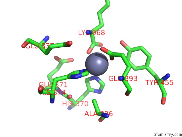
Mono view
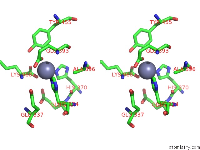
Stereo pair view
Mono view
Stereo pair view
A full contact list of Zinc with other atoms in the Zn binding
site number 1 of Crystal Structure of the Human Endoplasmic Reticulum Aminopeptidase 2 within 5.0Å range:
|
Zinc binding site 2 out of 2 in 3se6
Go back to
Zinc binding site 2 out
of 2 in the Crystal Structure of the Human Endoplasmic Reticulum Aminopeptidase 2
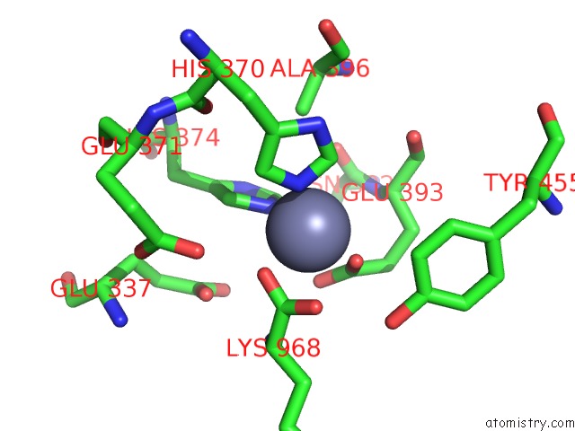
Mono view
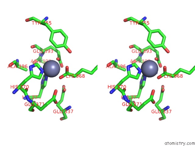
Stereo pair view
Mono view
Stereo pair view
A full contact list of Zinc with other atoms in the Zn binding
site number 2 of Crystal Structure of the Human Endoplasmic Reticulum Aminopeptidase 2 within 5.0Å range:
|
Reference:
J.R.Birtley,
E.Saridakis,
E.Stratikos,
I.M.Mavridis.
The Crystal Structure of Human Endoplasmic Reticulum Aminopeptidase 2 Reveals the Atomic Basis For Distinct Roles in Antigen Processing. Biochemistry V. 51 286 2012.
ISSN: ISSN 0006-2960
PubMed: 22106953
DOI: 10.1021/BI201230P
Page generated: Sat Oct 26 15:43:04 2024
ISSN: ISSN 0006-2960
PubMed: 22106953
DOI: 10.1021/BI201230P
Last articles
Zn in 9J0NZn in 9J0O
Zn in 9J0P
Zn in 9FJX
Zn in 9EKB
Zn in 9C0F
Zn in 9CAH
Zn in 9CH0
Zn in 9CH3
Zn in 9CH1