Zinc »
PDB 3rjn-3rxz »
3rtw »
Zinc in PDB 3rtw: Nitrowillardiine Bound to the Ligand Binding Domain of GLUA2
Protein crystallography data
The structure of Nitrowillardiine Bound to the Ligand Binding Domain of GLUA2, PDB code: 3rtw
was solved by
A.H.Ahmed,
R.E.Oswald,
with X-Ray Crystallography technique. A brief refinement statistics is given in the table below:
| Resolution Low / High (Å) | 28.57 / 2.10 |
| Space group | P 2 21 21 |
| Cell size a, b, c (Å), α, β, γ (°) | 48.077, 114.102, 165.048, 90.00, 90.00, 90.00 |
| R / Rfree (%) | 20.5 / 25.9 |
Zinc Binding Sites:
The binding sites of Zinc atom in the Nitrowillardiine Bound to the Ligand Binding Domain of GLUA2
(pdb code 3rtw). This binding sites where shown within
5.0 Angstroms radius around Zinc atom.
In total 5 binding sites of Zinc where determined in the Nitrowillardiine Bound to the Ligand Binding Domain of GLUA2, PDB code: 3rtw:
Jump to Zinc binding site number: 1; 2; 3; 4; 5;
In total 5 binding sites of Zinc where determined in the Nitrowillardiine Bound to the Ligand Binding Domain of GLUA2, PDB code: 3rtw:
Jump to Zinc binding site number: 1; 2; 3; 4; 5;
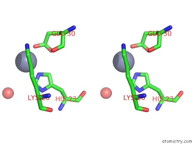
Zinc binding site 1 out of 5 in 3rtw
Go back to
Zinc binding site 1 out
of 5 in the Nitrowillardiine Bound to the Ligand Binding Domain of GLUA2
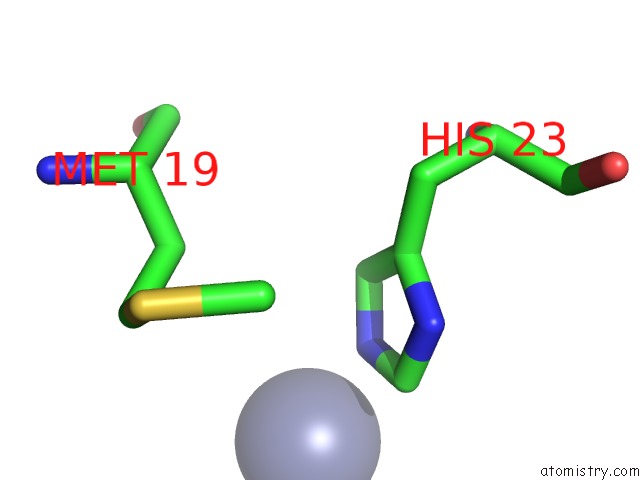
Mono view
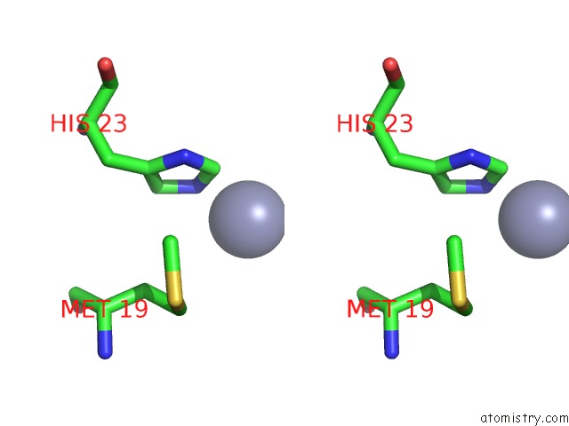
Stereo pair view
Mono view
Stereo pair view
A full contact list of Zinc with other atoms in the Zn binding
site number 1 of Nitrowillardiine Bound to the Ligand Binding Domain of GLUA2 within 5.0Å range:
|
Zinc binding site 2 out of 5 in 3rtw
Go back to
Zinc binding site 2 out
of 5 in the Nitrowillardiine Bound to the Ligand Binding Domain of GLUA2

Mono view
Stereo pair view
Mono view
Stereo pair view
A full contact list of Zinc with other atoms in the Zn binding
site number 2 of Nitrowillardiine Bound to the Ligand Binding Domain of GLUA2 within 5.0Å range:
|
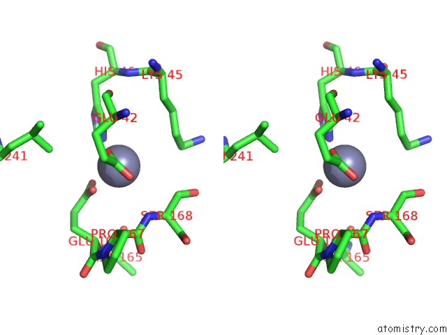
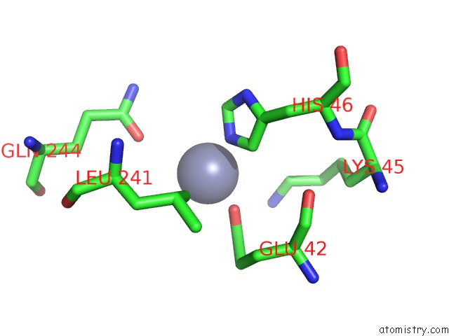
Zinc binding site 3 out of 5 in 3rtw
Go back to
Zinc binding site 3 out
of 5 in the Nitrowillardiine Bound to the Ligand Binding Domain of GLUA2
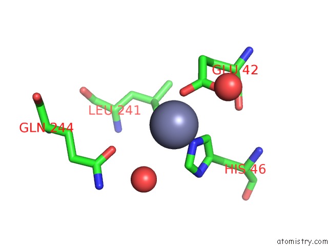
Mono view

Stereo pair view
Mono view
Stereo pair view
A full contact list of Zinc with other atoms in the Zn binding
site number 3 of Nitrowillardiine Bound to the Ligand Binding Domain of GLUA2 within 5.0Å range:
|
Zinc binding site 4 out of 5 in 3rtw
Go back to
Zinc binding site 4 out
of 5 in the Nitrowillardiine Bound to the Ligand Binding Domain of GLUA2

Mono view
Stereo pair view
Mono view
Stereo pair view
A full contact list of Zinc with other atoms in the Zn binding
site number 4 of Nitrowillardiine Bound to the Ligand Binding Domain of GLUA2 within 5.0Å range:
|
Zinc binding site 5 out of 5 in 3rtw
Go back to
Zinc binding site 5 out
of 5 in the Nitrowillardiine Bound to the Ligand Binding Domain of GLUA2
Mono view

Stereo pair view
Mono view
Stereo pair view
A full contact list of Zinc with other atoms in the Zn binding
site number 5 of Nitrowillardiine Bound to the Ligand Binding Domain of GLUA2 within 5.0Å range:
|
Reference:
K.Poon,
A.H.Ahmed,
L.M.Nowak,
R.E.Oswald.
Mechanisms of Modal Activation of GLUA3 Receptors. Mol.Pharmacol. V. 80 49 2011.
ISSN: ISSN 0026-895X
PubMed: 21464198
DOI: 10.1124/MOL.111.071688
Page generated: Sat Oct 26 15:04:47 2024
ISSN: ISSN 0026-895X
PubMed: 21464198
DOI: 10.1124/MOL.111.071688
Last articles
Zn in 9J0NZn in 9J0O
Zn in 9J0P
Zn in 9FJX
Zn in 9EKB
Zn in 9C0F
Zn in 9CAH
Zn in 9CH0
Zn in 9CH3
Zn in 9CH1