Zinc »
PDB 3rjw-3ryj »
3rn6 »
Zinc in PDB 3rn6: Crystal Structure of Cytosine Deaminase From Escherichia Coli Complexed with Zinc and Isoguanine
Enzymatic activity of Crystal Structure of Cytosine Deaminase From Escherichia Coli Complexed with Zinc and Isoguanine
All present enzymatic activity of Crystal Structure of Cytosine Deaminase From Escherichia Coli Complexed with Zinc and Isoguanine:
3.5.4.1;
3.5.4.1;
Protein crystallography data
The structure of Crystal Structure of Cytosine Deaminase From Escherichia Coli Complexed with Zinc and Isoguanine, PDB code: 3rn6
was solved by
A.A.Fedorov,
E.V.Fedorov,
D.S.Hitchcock,
F.M.Raushel,
S.C.Almo,
with X-Ray Crystallography technique. A brief refinement statistics is given in the table below:
| Resolution Low / High (Å) | 39.31 / 2.25 |
| Space group | H 3 2 |
| Cell size a, b, c (Å), α, β, γ (°) | 147.181, 147.181, 199.747, 90.00, 90.00, 120.00 |
| R / Rfree (%) | 20.7 / 24.3 |
Zinc Binding Sites:
The binding sites of Zinc atom in the Crystal Structure of Cytosine Deaminase From Escherichia Coli Complexed with Zinc and Isoguanine
(pdb code 3rn6). This binding sites where shown within
5.0 Angstroms radius around Zinc atom.
In total 2 binding sites of Zinc where determined in the Crystal Structure of Cytosine Deaminase From Escherichia Coli Complexed with Zinc and Isoguanine, PDB code: 3rn6:
Jump to Zinc binding site number: 1; 2;
In total 2 binding sites of Zinc where determined in the Crystal Structure of Cytosine Deaminase From Escherichia Coli Complexed with Zinc and Isoguanine, PDB code: 3rn6:
Jump to Zinc binding site number: 1; 2;
Zinc binding site 1 out of 2 in 3rn6
Go back to
Zinc binding site 1 out
of 2 in the Crystal Structure of Cytosine Deaminase From Escherichia Coli Complexed with Zinc and Isoguanine

Mono view
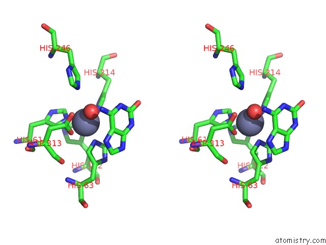
Stereo pair view
Mono view
Stereo pair view
A full contact list of Zinc with other atoms in the Zn binding
site number 1 of Crystal Structure of Cytosine Deaminase From Escherichia Coli Complexed with Zinc and Isoguanine within 5.0Å range:
|
Zinc binding site 2 out of 2 in 3rn6
Go back to
Zinc binding site 2 out
of 2 in the Crystal Structure of Cytosine Deaminase From Escherichia Coli Complexed with Zinc and Isoguanine
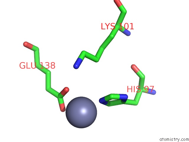
Mono view

Stereo pair view
Mono view
Stereo pair view
A full contact list of Zinc with other atoms in the Zn binding
site number 2 of Crystal Structure of Cytosine Deaminase From Escherichia Coli Complexed with Zinc and Isoguanine within 5.0Å range:
|
Reference:
D.S.Hitchcock,
A.A.Fedorov,
E.V.Fedorov,
L.J.Dangott,
S.C.Almo,
F.M.Raushel.
Rescue of the Orphan Enzyme Isoguanine Deaminase. Biochemistry V. 50 5555 2011.
ISSN: ISSN 0006-2960
PubMed: 21604715
DOI: 10.1021/BI200680Y
Page generated: Wed Aug 20 13:36:29 2025
ISSN: ISSN 0006-2960
PubMed: 21604715
DOI: 10.1021/BI200680Y
Last articles
Zn in 4IURZn in 4IUQ
Zn in 4IUP
Zn in 4IUM
Zn in 4IQJ
Zn in 4IUE
Zn in 4ITP
Zn in 4IS1
Zn in 4ITO
Zn in 4IQR