Zinc »
PDB 3r0d-3rja »
3rfr »
Zinc in PDB 3rfr: Crystal Structure of Particulate Methane Monooxygenase (Pmmo) From Methylocystis Sp. Strain M
Protein crystallography data
The structure of Crystal Structure of Particulate Methane Monooxygenase (Pmmo) From Methylocystis Sp. Strain M, PDB code: 3rfr
was solved by
S.M.Smith,
A.C.Rosenzweig,
with X-Ray Crystallography technique. A brief refinement statistics is given in the table below:
| Resolution Low / High (Å) | 45.79 / 2.68 |
| Space group | P 21 21 21 |
| Cell size a, b, c (Å), α, β, γ (°) | 107.718, 178.310, 183.147, 90.00, 90.00, 90.00 |
| R / Rfree (%) | 24.9 / 28.1 |
Other elements in 3rfr:
The structure of Crystal Structure of Particulate Methane Monooxygenase (Pmmo) From Methylocystis Sp. Strain M also contains other interesting chemical elements:
| Copper | (Cu) | 4 atoms |
Zinc Binding Sites:
The binding sites of Zinc atom in the Crystal Structure of Particulate Methane Monooxygenase (Pmmo) From Methylocystis Sp. Strain M
(pdb code 3rfr). This binding sites where shown within
5.0 Angstroms radius around Zinc atom.
In total 9 binding sites of Zinc where determined in the Crystal Structure of Particulate Methane Monooxygenase (Pmmo) From Methylocystis Sp. Strain M, PDB code: 3rfr:
Jump to Zinc binding site number: 1; 2; 3; 4; 5; 6; 7; 8; 9;
In total 9 binding sites of Zinc where determined in the Crystal Structure of Particulate Methane Monooxygenase (Pmmo) From Methylocystis Sp. Strain M, PDB code: 3rfr:
Jump to Zinc binding site number: 1; 2; 3; 4; 5; 6; 7; 8; 9;
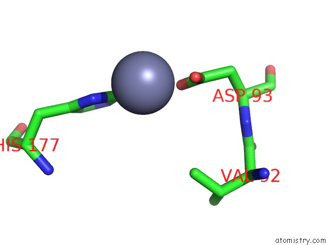
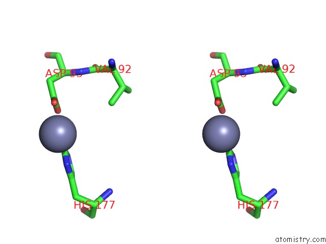
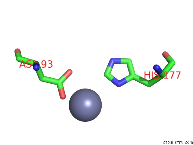
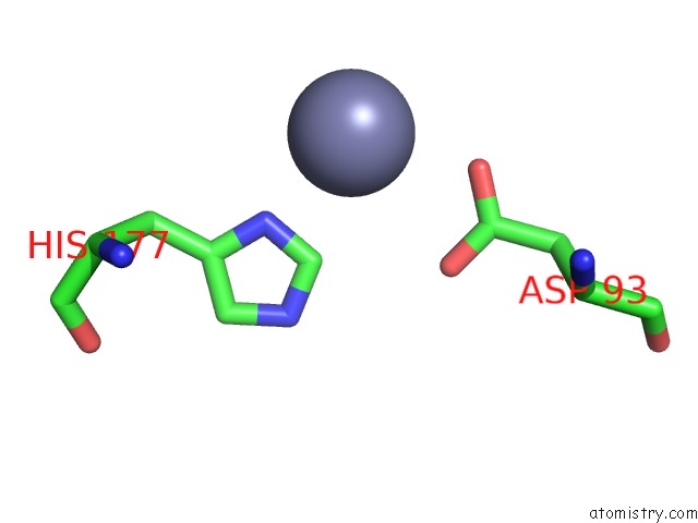
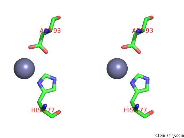
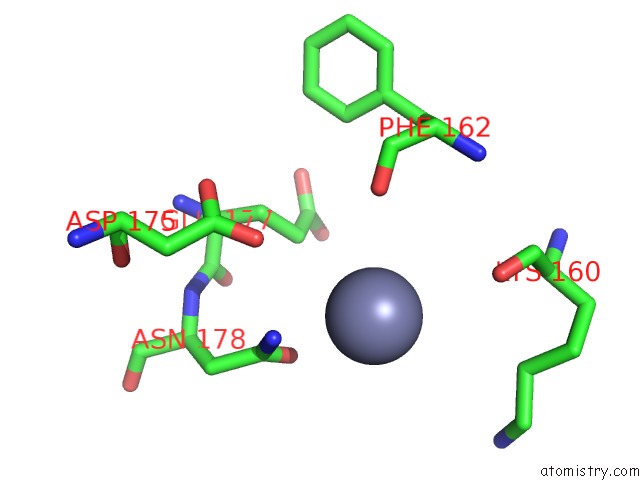
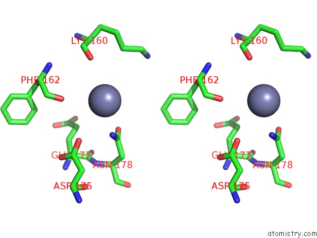
Zinc binding site 1 out of 9 in 3rfr
Go back to
Zinc binding site 1 out
of 9 in the Crystal Structure of Particulate Methane Monooxygenase (Pmmo) From Methylocystis Sp. Strain M
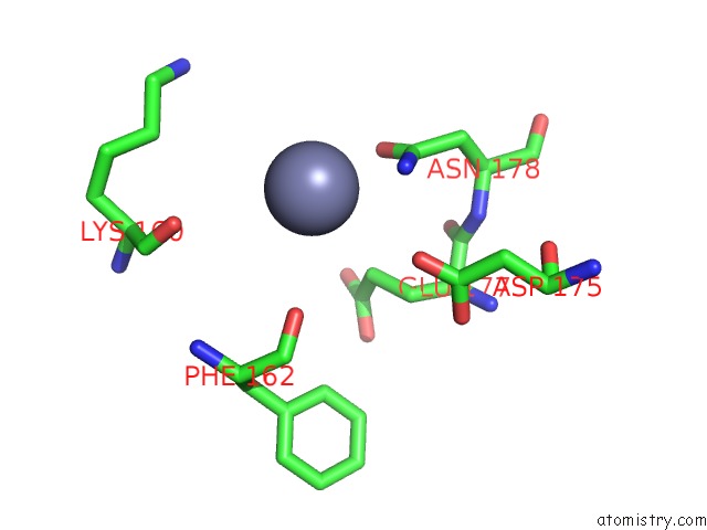
Mono view
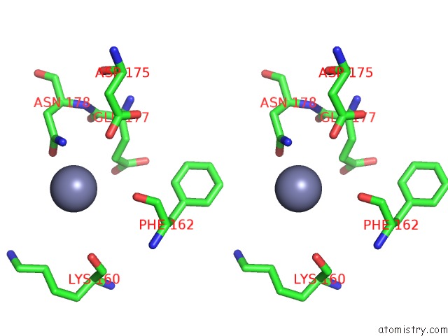
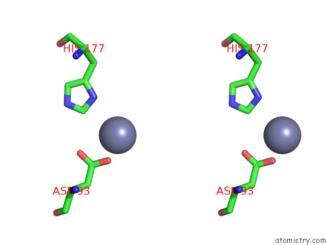
Stereo pair view
Mono view
Stereo pair view
A full contact list of Zinc with other atoms in the Zn binding
site number 1 of Crystal Structure of Particulate Methane Monooxygenase (Pmmo) From Methylocystis Sp. Strain M within 5.0Å range:
|
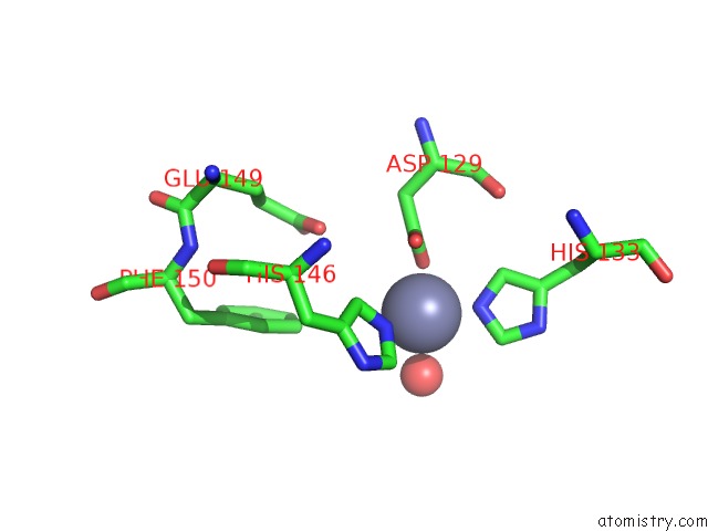
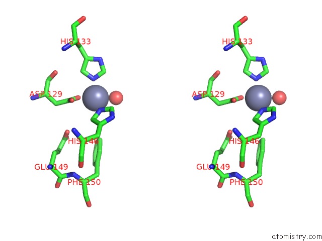
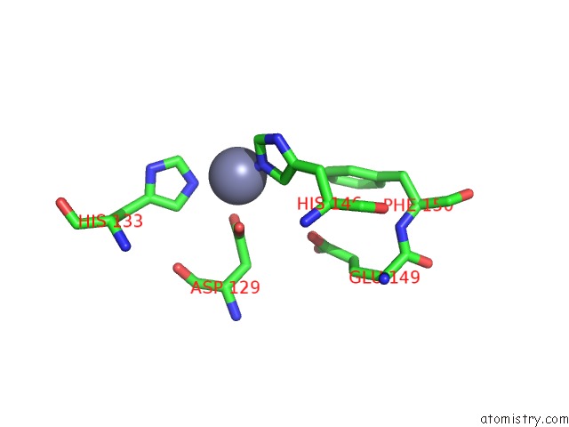
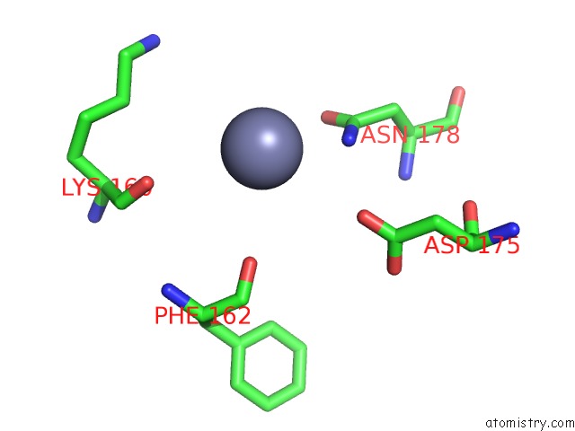
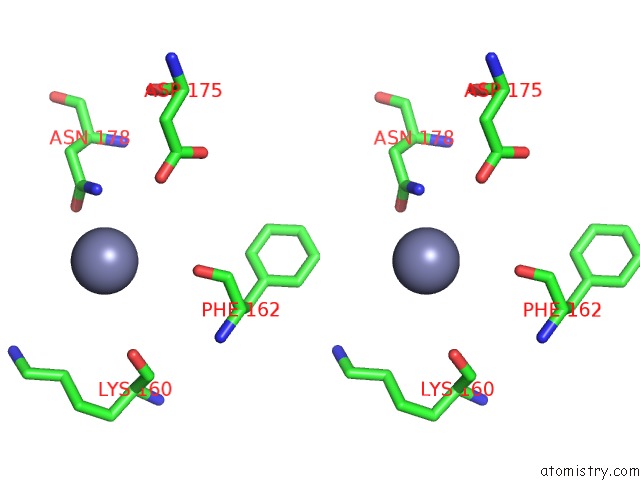
Zinc binding site 2 out of 9 in 3rfr
Go back to
Zinc binding site 2 out
of 9 in the Crystal Structure of Particulate Methane Monooxygenase (Pmmo) From Methylocystis Sp. Strain M
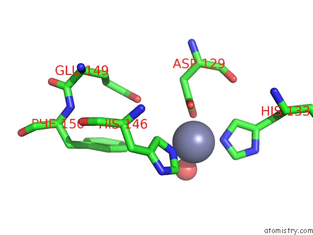
Mono view
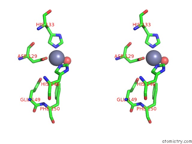
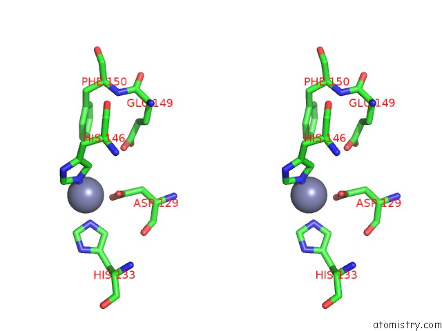
Stereo pair view
Mono view
Stereo pair view
A full contact list of Zinc with other atoms in the Zn binding
site number 2 of Crystal Structure of Particulate Methane Monooxygenase (Pmmo) From Methylocystis Sp. Strain M within 5.0Å range:
|
Zinc binding site 3 out of 9 in 3rfr
Go back to
Zinc binding site 3 out
of 9 in the Crystal Structure of Particulate Methane Monooxygenase (Pmmo) From Methylocystis Sp. Strain M
Mono view
Stereo pair view
Mono view
Stereo pair view
A full contact list of Zinc with other atoms in the Zn binding
site number 3 of Crystal Structure of Particulate Methane Monooxygenase (Pmmo) From Methylocystis Sp. Strain M within 5.0Å range:
|
Zinc binding site 4 out of 9 in 3rfr
Go back to
Zinc binding site 4 out
of 9 in the Crystal Structure of Particulate Methane Monooxygenase (Pmmo) From Methylocystis Sp. Strain M
Mono view
Stereo pair view
Mono view
Stereo pair view
A full contact list of Zinc with other atoms in the Zn binding
site number 4 of Crystal Structure of Particulate Methane Monooxygenase (Pmmo) From Methylocystis Sp. Strain M within 5.0Å range:
|
Zinc binding site 5 out of 9 in 3rfr
Go back to
Zinc binding site 5 out
of 9 in the Crystal Structure of Particulate Methane Monooxygenase (Pmmo) From Methylocystis Sp. Strain M
Mono view
Stereo pair view
Mono view
Stereo pair view
A full contact list of Zinc with other atoms in the Zn binding
site number 5 of Crystal Structure of Particulate Methane Monooxygenase (Pmmo) From Methylocystis Sp. Strain M within 5.0Å range:
|
Zinc binding site 6 out of 9 in 3rfr
Go back to
Zinc binding site 6 out
of 9 in the Crystal Structure of Particulate Methane Monooxygenase (Pmmo) From Methylocystis Sp. Strain M
Mono view
Stereo pair view
Mono view
Stereo pair view
A full contact list of Zinc with other atoms in the Zn binding
site number 6 of Crystal Structure of Particulate Methane Monooxygenase (Pmmo) From Methylocystis Sp. Strain M within 5.0Å range:
|
Zinc binding site 7 out of 9 in 3rfr
Go back to
Zinc binding site 7 out
of 9 in the Crystal Structure of Particulate Methane Monooxygenase (Pmmo) From Methylocystis Sp. Strain M
Mono view
Stereo pair view
Mono view
Stereo pair view
A full contact list of Zinc with other atoms in the Zn binding
site number 7 of Crystal Structure of Particulate Methane Monooxygenase (Pmmo) From Methylocystis Sp. Strain M within 5.0Å range:
|
Zinc binding site 8 out of 9 in 3rfr
Go back to
Zinc binding site 8 out
of 9 in the Crystal Structure of Particulate Methane Monooxygenase (Pmmo) From Methylocystis Sp. Strain M
Mono view
Stereo pair view
Mono view
Stereo pair view
A full contact list of Zinc with other atoms in the Zn binding
site number 8 of Crystal Structure of Particulate Methane Monooxygenase (Pmmo) From Methylocystis Sp. Strain M within 5.0Å range:
|
Zinc binding site 9 out of 9 in 3rfr
Go back to
Zinc binding site 9 out
of 9 in the Crystal Structure of Particulate Methane Monooxygenase (Pmmo) From Methylocystis Sp. Strain M
Mono view
Stereo pair view
Mono view
Stereo pair view
A full contact list of Zinc with other atoms in the Zn binding
site number 9 of Crystal Structure of Particulate Methane Monooxygenase (Pmmo) From Methylocystis Sp. Strain M within 5.0Å range:
|
Reference:
S.M.Smith,
S.Rawat,
J.Telser,
B.M.Hoffman,
T.L.Stemmler,
A.C.Rosenzweig.
Crystal Structure and Characterization of Particulate Methane Monooxygenase From Methylocystis Species Strain M. Biochemistry V. 50 10231 2011.
ISSN: ISSN 0006-2960
PubMed: 22013879
DOI: 10.1021/BI200801Z
Page generated: Sat Oct 26 14:45:37 2024
ISSN: ISSN 0006-2960
PubMed: 22013879
DOI: 10.1021/BI200801Z
Last articles
Zn in 9J0NZn in 9J0O
Zn in 9J0P
Zn in 9FJX
Zn in 9EKB
Zn in 9C0F
Zn in 9CAH
Zn in 9CH0
Zn in 9CH3
Zn in 9CH1