Zinc »
PDB 3p4v-3phd »
3p76 »
Zinc in PDB 3p76: X-Ray Crystal Structure of Aquifex Aeolicus Lpxc Complexed SCH1379777
Protein crystallography data
The structure of X-Ray Crystal Structure of Aquifex Aeolicus Lpxc Complexed SCH1379777, PDB code: 3p76
was solved by
P.Orth,
with X-Ray Crystallography technique. A brief refinement statistics is given in the table below:
| Resolution Low / High (Å) | 29.44 / 1.93 |
| Space group | P 61 |
| Cell size a, b, c (Å), α, β, γ (°) | 65.592, 65.592, 133.588, 90.00, 90.00, 120.00 |
| R / Rfree (%) | 16.8 / 18.5 |
Zinc Binding Sites:
The binding sites of Zinc atom in the X-Ray Crystal Structure of Aquifex Aeolicus Lpxc Complexed SCH1379777
(pdb code 3p76). This binding sites where shown within
5.0 Angstroms radius around Zinc atom.
In total 2 binding sites of Zinc where determined in the X-Ray Crystal Structure of Aquifex Aeolicus Lpxc Complexed SCH1379777, PDB code: 3p76:
Jump to Zinc binding site number: 1; 2;
In total 2 binding sites of Zinc where determined in the X-Ray Crystal Structure of Aquifex Aeolicus Lpxc Complexed SCH1379777, PDB code: 3p76:
Jump to Zinc binding site number: 1; 2;
Zinc binding site 1 out of 2 in 3p76
Go back to
Zinc binding site 1 out
of 2 in the X-Ray Crystal Structure of Aquifex Aeolicus Lpxc Complexed SCH1379777
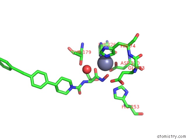
Mono view
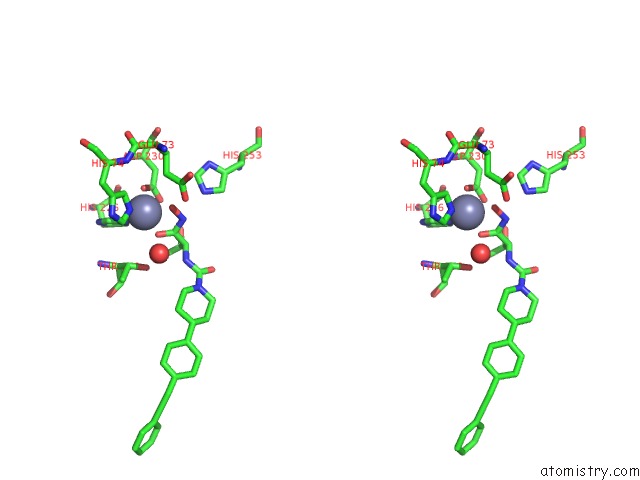
Stereo pair view
Mono view
Stereo pair view
A full contact list of Zinc with other atoms in the Zn binding
site number 1 of X-Ray Crystal Structure of Aquifex Aeolicus Lpxc Complexed SCH1379777 within 5.0Å range:
|
Zinc binding site 2 out of 2 in 3p76
Go back to
Zinc binding site 2 out
of 2 in the X-Ray Crystal Structure of Aquifex Aeolicus Lpxc Complexed SCH1379777
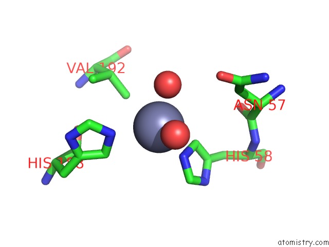
Mono view
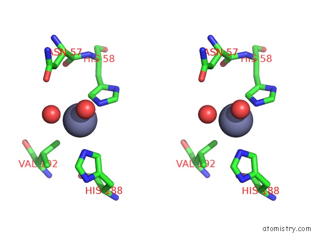
Stereo pair view
Mono view
Stereo pair view
A full contact list of Zinc with other atoms in the Zn binding
site number 2 of X-Ray Crystal Structure of Aquifex Aeolicus Lpxc Complexed SCH1379777 within 5.0Å range:
|
Reference:
U.Faruk Mansoor,
D.Vitharana,
P.A.Reddy,
D.L.Daubaras,
P.Mcnicholas,
P.Orth,
T.Black,
M.Arshad Siddiqui.
Design and Synthesis of Potent Gram-Negative Specific Lpxc Inhibitors. Bioorg.Med.Chem.Lett. V. 21 1155 2011.
ISSN: ISSN 0960-894X
PubMed: 21273067
DOI: 10.1016/J.BMCL.2010.12.111
Page generated: Sat Oct 26 11:20:32 2024
ISSN: ISSN 0960-894X
PubMed: 21273067
DOI: 10.1016/J.BMCL.2010.12.111
Last articles
Zn in 9J0NZn in 9J0O
Zn in 9J0P
Zn in 9FJX
Zn in 9EKB
Zn in 9C0F
Zn in 9CAH
Zn in 9CH0
Zn in 9CH3
Zn in 9CH1