Zinc »
PDB 3oxk-3p44 »
3oxk »
Zinc in PDB 3oxk: Crystal Structure of A Histidine Triad Family Protein From Entamoeba Histolytica, Bound to Gmp
Protein crystallography data
The structure of Crystal Structure of A Histidine Triad Family Protein From Entamoeba Histolytica, Bound to Gmp, PDB code: 3oxk
was solved by
Seattle Structural Genomics Center For Infectious Disease (Ssgcid),
with X-Ray Crystallography technique. A brief refinement statistics is given in the table below:
| Resolution Low / High (Å) | 27.77 / 1.55 |
| Space group | C 2 2 21 |
| Cell size a, b, c (Å), α, β, γ (°) | 53.660, 60.860, 67.930, 90.00, 90.00, 90.00 |
| R / Rfree (%) | 15.7 / 20.8 |
Zinc Binding Sites:
The binding sites of Zinc atom in the Crystal Structure of A Histidine Triad Family Protein From Entamoeba Histolytica, Bound to Gmp
(pdb code 3oxk). This binding sites where shown within
5.0 Angstroms radius around Zinc atom.
In total only one binding site of Zinc was determined in the Crystal Structure of A Histidine Triad Family Protein From Entamoeba Histolytica, Bound to Gmp, PDB code: 3oxk:
In total only one binding site of Zinc was determined in the Crystal Structure of A Histidine Triad Family Protein From Entamoeba Histolytica, Bound to Gmp, PDB code: 3oxk:
Zinc binding site 1 out of 1 in 3oxk
Go back to
Zinc binding site 1 out
of 1 in the Crystal Structure of A Histidine Triad Family Protein From Entamoeba Histolytica, Bound to Gmp
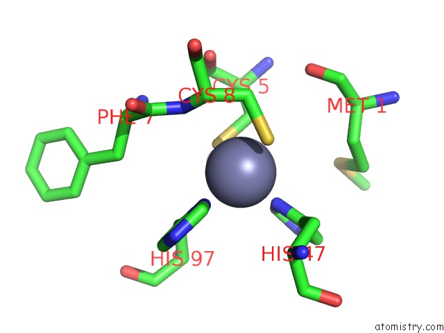
Mono view
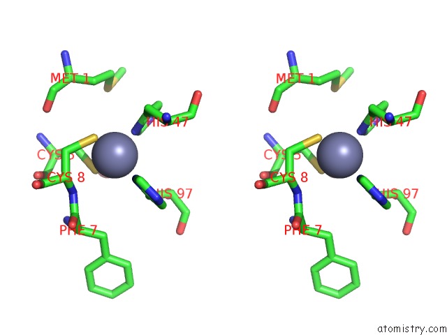
Stereo pair view
Mono view
Stereo pair view
A full contact list of Zinc with other atoms in the Zn binding
site number 1 of Crystal Structure of A Histidine Triad Family Protein From Entamoeba Histolytica, Bound to Gmp within 5.0Å range:
|
Reference:
D.D.Lorimer,
R.Choi,
A.Abramov,
S.Nakazawa Hewitt,
A.S.Gardberg,
W.C.Van Voorhis,
B.L.Staker,
P.J.Myler,
T.E.Edwards.
Structures of A Histidine Triad Family Protein From Entamoeba Histolytica Bound to Sulfate, Amp and Gmp. Acta Crystallogr F Struct V. 71 572 2015BIOL Commun.
PubMed: 25945711
DOI: 10.1107/S2053230X1500237X
Page generated: Sat Oct 26 11:10:41 2024
PubMed: 25945711
DOI: 10.1107/S2053230X1500237X
Last articles
Zn in 9J0NZn in 9J0O
Zn in 9J0P
Zn in 9FJX
Zn in 9EKB
Zn in 9C0F
Zn in 9CAH
Zn in 9CH0
Zn in 9CH3
Zn in 9CH1