Zinc »
PDB 3okl-3oxg »
3ooi »
Zinc in PDB 3ooi: Crystal Structure of Human Histone-Lysine N-Methyltransferase NSD1 Set Domain in Complex with S-Adenosyl-L-Methionine
Enzymatic activity of Crystal Structure of Human Histone-Lysine N-Methyltransferase NSD1 Set Domain in Complex with S-Adenosyl-L-Methionine
All present enzymatic activity of Crystal Structure of Human Histone-Lysine N-Methyltransferase NSD1 Set Domain in Complex with S-Adenosyl-L-Methionine:
2.1.1.43;
2.1.1.43;
Protein crystallography data
The structure of Crystal Structure of Human Histone-Lysine N-Methyltransferase NSD1 Set Domain in Complex with S-Adenosyl-L-Methionine, PDB code: 3ooi
was solved by
Q.Qiao,
M.Wang,
R.M.Xu,
with X-Ray Crystallography technique. A brief refinement statistics is given in the table below:
| Resolution Low / High (Å) | 27.61 / 1.75 |
| Space group | P 21 21 21 |
| Cell size a, b, c (Å), α, β, γ (°) | 65.875, 67.751, 69.086, 90.00, 90.00, 90.00 |
| R / Rfree (%) | 16.4 / 18.6 |
Zinc Binding Sites:
The binding sites of Zinc atom in the Crystal Structure of Human Histone-Lysine N-Methyltransferase NSD1 Set Domain in Complex with S-Adenosyl-L-Methionine
(pdb code 3ooi). This binding sites where shown within
5.0 Angstroms radius around Zinc atom.
In total 3 binding sites of Zinc where determined in the Crystal Structure of Human Histone-Lysine N-Methyltransferase NSD1 Set Domain in Complex with S-Adenosyl-L-Methionine, PDB code: 3ooi:
Jump to Zinc binding site number: 1; 2; 3;
In total 3 binding sites of Zinc where determined in the Crystal Structure of Human Histone-Lysine N-Methyltransferase NSD1 Set Domain in Complex with S-Adenosyl-L-Methionine, PDB code: 3ooi:
Jump to Zinc binding site number: 1; 2; 3;
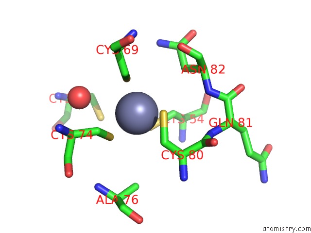
Zinc binding site 1 out of 3 in 3ooi
Go back to
Zinc binding site 1 out
of 3 in the Crystal Structure of Human Histone-Lysine N-Methyltransferase NSD1 Set Domain in Complex with S-Adenosyl-L-Methionine
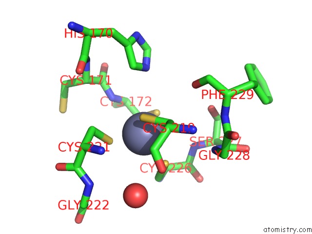
Mono view
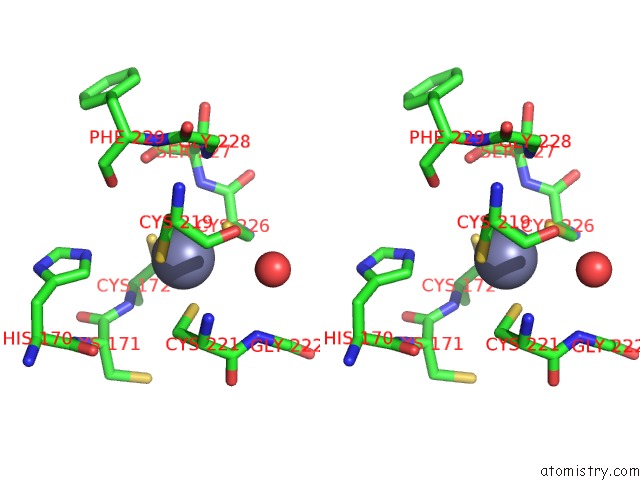
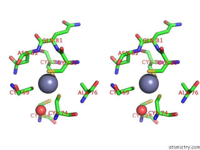
Stereo pair view
Mono view
Stereo pair view
A full contact list of Zinc with other atoms in the Zn binding
site number 1 of Crystal Structure of Human Histone-Lysine N-Methyltransferase NSD1 Set Domain in Complex with S-Adenosyl-L-Methionine within 5.0Å range:
|
Zinc binding site 2 out of 3 in 3ooi
Go back to
Zinc binding site 2 out
of 3 in the Crystal Structure of Human Histone-Lysine N-Methyltransferase NSD1 Set Domain in Complex with S-Adenosyl-L-Methionine
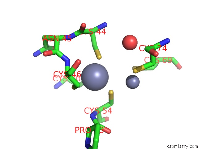
Mono view
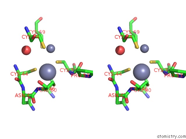
Stereo pair view
Mono view
Stereo pair view
A full contact list of Zinc with other atoms in the Zn binding
site number 2 of Crystal Structure of Human Histone-Lysine N-Methyltransferase NSD1 Set Domain in Complex with S-Adenosyl-L-Methionine within 5.0Å range:
|
Zinc binding site 3 out of 3 in 3ooi
Go back to
Zinc binding site 3 out
of 3 in the Crystal Structure of Human Histone-Lysine N-Methyltransferase NSD1 Set Domain in Complex with S-Adenosyl-L-Methionine
Mono view
Stereo pair view
Mono view
Stereo pair view
A full contact list of Zinc with other atoms in the Zn binding
site number 3 of Crystal Structure of Human Histone-Lysine N-Methyltransferase NSD1 Set Domain in Complex with S-Adenosyl-L-Methionine within 5.0Å range:
|
Reference:
Q.Qiao,
Y.Li,
Z.Chen,
M.Wang,
D.Reinberg,
R.M.Xu.
The Structure of NSD1 Reveals An Autoregulatory Mechanism Underlying Histone H3K36 Methylation J.Biol.Chem. V. 286 8361 2010.
ISSN: ISSN 0021-9258
PubMed: 21196496
DOI: 10.1074/JBC.M110.204115
Page generated: Sat Oct 26 11:03:06 2024
ISSN: ISSN 0021-9258
PubMed: 21196496
DOI: 10.1074/JBC.M110.204115
Last articles
Zn in 9J0NZn in 9J0O
Zn in 9J0P
Zn in 9FJX
Zn in 9EKB
Zn in 9C0F
Zn in 9CAH
Zn in 9CH0
Zn in 9CH3
Zn in 9CH1