Zinc »
PDB 3lta-3m1m »
3lta »
Zinc in PDB 3lta: Crystal Structure of A Non-Biological Atp Binding Protein with A Tyr- Phe Mutation Within the Ligand Binding Domain
Protein crystallography data
The structure of Crystal Structure of A Non-Biological Atp Binding Protein with A Tyr- Phe Mutation Within the Ligand Binding Domain, PDB code: 3lta
was solved by
C.R.Simmons,
C.L.Magee,
J.P.Allen,
J.C.Chaput,
with X-Ray Crystallography technique. A brief refinement statistics is given in the table below:
| Resolution Low / High (Å) | 41.42 / 2.70 |
| Space group | P 32 2 1 |
| Cell size a, b, c (Å), α, β, γ (°) | 71.107, 71.107, 55.935, 90.00, 90.00, 120.00 |
| R / Rfree (%) | 20.2 / 23.8 |
Other elements in 3lta:
The structure of Crystal Structure of A Non-Biological Atp Binding Protein with A Tyr- Phe Mutation Within the Ligand Binding Domain also contains other interesting chemical elements:
| Chlorine | (Cl) | 1 atom |
Zinc Binding Sites:
The binding sites of Zinc atom in the Crystal Structure of A Non-Biological Atp Binding Protein with A Tyr- Phe Mutation Within the Ligand Binding Domain
(pdb code 3lta). This binding sites where shown within
5.0 Angstroms radius around Zinc atom.
In total only one binding site of Zinc was determined in the Crystal Structure of A Non-Biological Atp Binding Protein with A Tyr- Phe Mutation Within the Ligand Binding Domain, PDB code: 3lta:
In total only one binding site of Zinc was determined in the Crystal Structure of A Non-Biological Atp Binding Protein with A Tyr- Phe Mutation Within the Ligand Binding Domain, PDB code: 3lta:
Zinc binding site 1 out of 1 in 3lta
Go back to
Zinc binding site 1 out
of 1 in the Crystal Structure of A Non-Biological Atp Binding Protein with A Tyr- Phe Mutation Within the Ligand Binding Domain
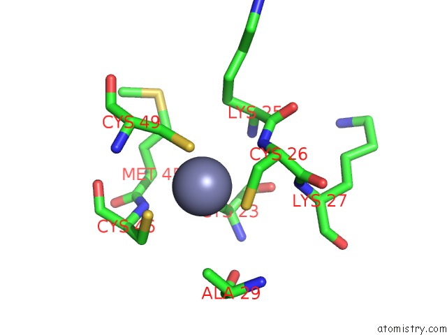
Mono view
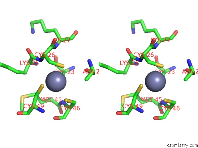
Stereo pair view
Mono view
Stereo pair view
A full contact list of Zinc with other atoms in the Zn binding
site number 1 of Crystal Structure of A Non-Biological Atp Binding Protein with A Tyr- Phe Mutation Within the Ligand Binding Domain within 5.0Å range:
|
Reference:
C.R.Simmons,
C.L.Magee,
D.A.Smith,
L.Lauman,
J.C.Chaput,
J.P.Allen.
Three-Dimensional Structures Reveal Multiple Adp/Atp Binding Modes For A Synthetic Class of Artificial Proteins. Biochemistry V. 49 8689 2010.
ISSN: ISSN 0006-2960
PubMed: 20822107
DOI: 10.1021/BI100398P
Page generated: Sat Oct 26 08:52:59 2024
ISSN: ISSN 0006-2960
PubMed: 20822107
DOI: 10.1021/BI100398P
Last articles
Zn in 9J0NZn in 9J0O
Zn in 9J0P
Zn in 9FJX
Zn in 9EKB
Zn in 9C0F
Zn in 9CAH
Zn in 9CH0
Zn in 9CH3
Zn in 9CH1